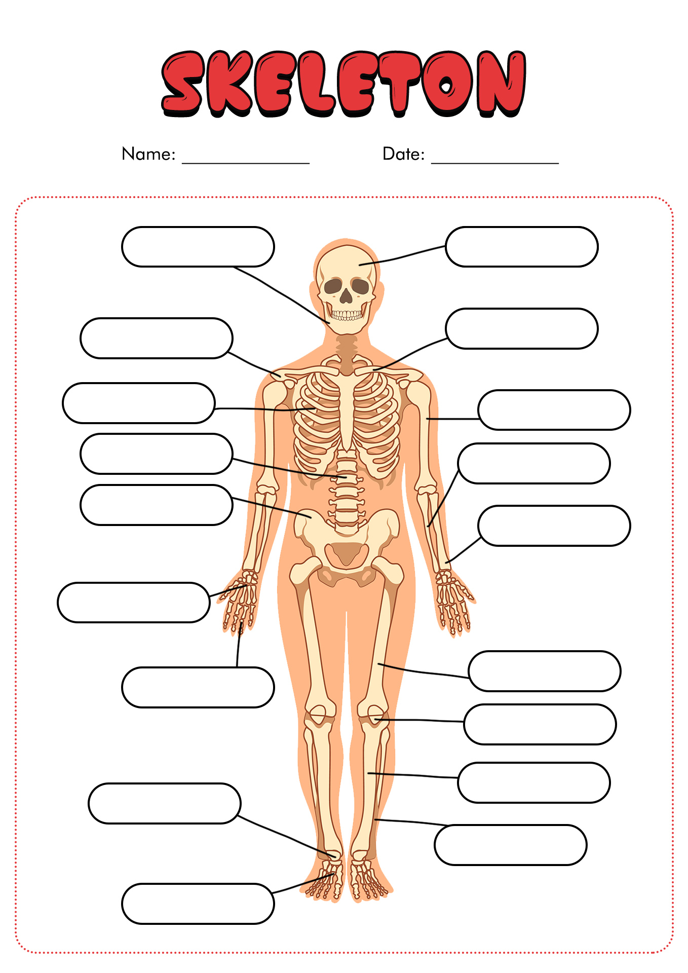
44 simple skeleton diagram to label
Drawing Feet | Muddy Colors Thank the feet for that arch, that we need to put the leg over and not behind. The ankle bones are the tibia and the fibula together, and combined they form a wrench like structure that holds the foot bones in place as the pivot point for the foot rotation. The bones are side by side medially and laterally, not in front and behind. List of skeletal muscles of the human body - Wikipedia The terms "artery" and "nerve" are both used when these structures are mentioned. Contents 1 Head 1.1 Forehead/eyelid 1.2 Extraocular muscles 1.3 Ear 1.4 Nose 1.5 Mouth 1.6 Mastication 1.7 Tongue 1.7.1 Extrinsic muscle 1.7.2 Intrinsic 1.8 Soft palate 1.9 Pharynx 1.10 Larynx 2 Neck 2.1 Clavicular 2.2 Suprahyoid 2.3 Infrahyoid 2.4 Neck 2.4.1 Anterior
Binocular Microscope Anatomy - Parts and Functions with a Labeled Diagram It is essential to know the binocular microscope anatomy for the first-year veterinary student before starting histology learning. This article might help you know different parts of the binocular compound microscope with a labeled diagram. Most first-year students don't know how perfectly set up a slide under the light microscope.

Simple skeleton diagram to label
Parts and Components of Human Ear and Their Functions - MD-Health.com In addition to helping the body take in auditory messages, the ear helps to maintain a proper head position. The fluid in the ear also helps the body maintain a sense of balance so the body can maintain proper posture and coordination. There are three major parts of the ear, the outer, middle and inner ear. Each contains several parts that are ... Neck Space Anatomy | SpringerLink In diagrams, these layers are red (superficial), blue (middle), and yellow (deep). The spaces created by this arrangement (Fig. 6) are; Masticator. Parotid. Pharyngeal mucosal space. Carotid. Retropharyngeal and danger spaces. Parapharyngeal. Perivertebral. Fig. 6 Axial T1W MRI suprahyoid neck and corresponding line diagram. Appendicular Skeleton: Bones List, Diagram & More - Embibe The Human Skeletal system consists of a framework of bones and a few cartilages. This system has a significant role in the movement shown by the body. (Source: NCERT) The skeletal system of humans can be basically divided into two parts (i) the axial skeleton and (ii) the appendicular skeleton.
Simple skeleton diagram to label. Anatomy Coloring Page - sofia-blogbright.blogspot.com The Anatomy Coloring Book 4th Edition As one of the most popular anatomy coloring books of all time and its easy to see why. They have hard rounded shells covered with sharp movable spines. Please check the lesson plans to see what you missed and for make-up work. The tail which measures about 1 meter in length is slightly thick with fringes. The Gods of Galas Porras-Kim | The Nation In lieu of tombstones and wall labels, ... the name given to an 11,500-year-old skeleton, among the earliest intact human remains. ... Porras-Kim, in a deceptively simple manner, points to the ... Kidney Structures and Functions Explained (with Picture and Video) Each apex of the renal pyramid is connected to a minor calyx, a hollow collecting tube for urine. These minor calyces merge and form three major calyces that also merge into the renal pelvis at the hilus of the kidney. From here, urine drains into the larger ureter. Double Helix - Genome.gov Double helix, as related to genomics, is a term used to describe the physical structure of DNA. A DNA molecule is made up of two linked strands that wind around each other to resemble a twisted ladder in a helix-like shape. Each strand has a backbone made of alternating sugar (deoxyribose) and phosphate groups.
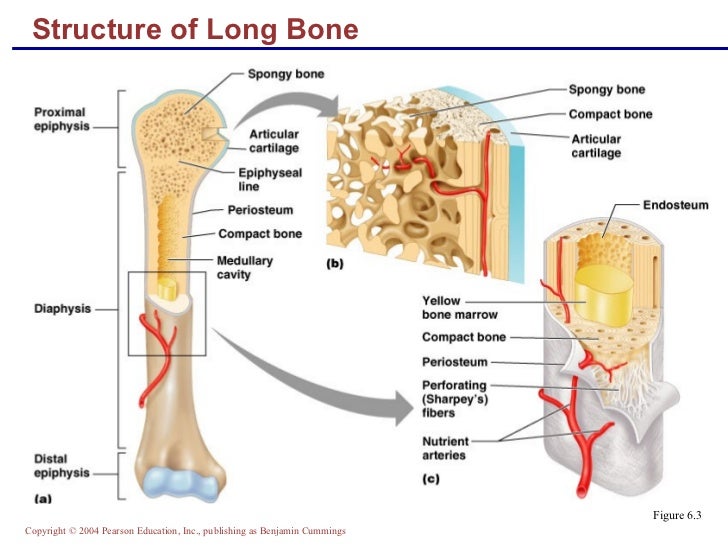
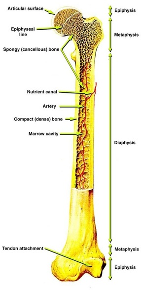
What are the different parts and functions of an otoscope? References. Answer. An otoscope consists of a head and a handle and is used to examine the external auditory canal (EAC), the tympanic membrane, and the middle ear. A magnifying lens enhances the ... Axial Skeleton Anatomy: Diagram, Definition, Functions - Embibe The axial skeleton is the section of a vertebrate's skeleton that comprises the head and trunk bones. The human skeleton is made up of 80 bones and is divided into six sections: the skull (22 bones), middle ear ossicles, hyoid bone, rib cage, sternum, and spinal column. The axial and appendicular skeletons combine to produce the entire skeleton. Stage Gate process: How to prevent project risk - asana.com Basic For simple task and project management. Free for teams up to 15. ... Below is an example of the Stage Gate process, with each stage and gate labeled. The project must go through each gate before moving to the next project phase. ... Workflow diagram: Symbols, uses, and examples. Article Das Agile Manifesto: Definition, Prinzipien und ... Organs of the body | Their Locations and Internal Functions Below, we will see essential organs present in different human anatomy locations. List of Organs of the body. Organs are the structures formed by a group of tissues. The human body organs of different types and can be grouped into sections like. Digestion: Esophagus, Stomach, liver, pancreas, small intestine, large intestine, rectum, anus and ...
WHMIS 2015 - Pictograms : OSH Answers Pictograms are graphic images that immediately show the user of a hazardous product what type of hazard is present. With a quick glance, you can see, for example, that the product is flammable, or if it might be a health hazard. Most pictograms have a distinctive red "square set on one of its points" border. A Guide to Different Ear Piercing Types and Their Positions Piercing the cartilage above the earlobe and the anti-tragus on the inside of the ear. Piercing of the thick fold of cartilage on the upper inside of the ear. A piercing of the inner cartilage half-way down the outer rim of the ear. A piercing between rook and the ear canal. Any piercing on the outer rim of the ear. Data structures for effective Python applications The following diagram shows the different types of Python data structures both built-in and user-defined. In the following section of this tutorial, you will use the knowledge you have just gained to create a simple API that will allow you to store, manipulate, and retrieve data from data structures. API flow diagram and storage Machine learning for contour classification in TG-263 noncompliant ... Of the 546 data sets, all patients contoured before TG-263 were labeled with an in-house-standardized nomenclature; patients after TG-263 were labeled with a TG-263 compliant scheme. Each CT image had a size of 512 × 512 voxels and images were not preprocessed to match voxel dimensions, with a uniform slice thickness of 2.5 mm.
100 YEARS TEACHING CHILDREN: LABEL THE SKELETON 2
Human brain - Wikipedia Each hemisphere is conventionally divided into four main lobes; the frontal lobe, parietal lobe, temporal lobe, and occipital lobe, named according to the skull bones that overlie them. [9] Each lobe is associated with one or two specialised functions though there is some functional overlap between them. [18]
What does "bone structure" mean? | Socratic
Circulatory System Diagram - New Health Advisor There are different types of circulatory system diagrams; some have labels while others don't. The color blue stands for deoxygenated blood while red stands for blood which is oxygenated. Below you'll see diagram specified to the heart, as well as circulatory system diagram of the whole body: How Does the Human Circulatory System Work? 1. Heart

Brain Jack Image: Brain Diagram
Positions and Functions of the Four Brain Lobes - MD-Health.com The occipital lobe, the smallest of the four lobes of the brain, is located near the posterior region of the cerebral cortex, near the back of the skull. The occipital lobe is the primary visual processing center of the brain. Here are some other functions of the occipital lobe: Visual-spatial processing. Movement and color recognition.

skeleton label worksheet with answer key | Anatomy coloring book, Human ...
Simple epithelium: Location, function, structure | Kenhub Simple squamous epithelium 1/4 Simple epithelium has only one cell layer where every cell is in direct contact with the underlying basement membrane. Generally, this type of epithelium is found inside the body probably due to the fragile nature and forms the lining of the body cavities, blood and lymph vessels, heart and respiratory system.
Skeletal System - My Future Depends on Me
Gram Stain Technique (Theory) : Microbiology Virtual Lab I ... 2) Addition of Gram's Iodine. Iodine (I - or I3 -) acts as a mordant and as a trapping agent. A mordant is a substance that increases the affinity of the cell wall for a stain by binding to the primary stain, thus forming an insoluble complex which gets trapped in the cell wall.
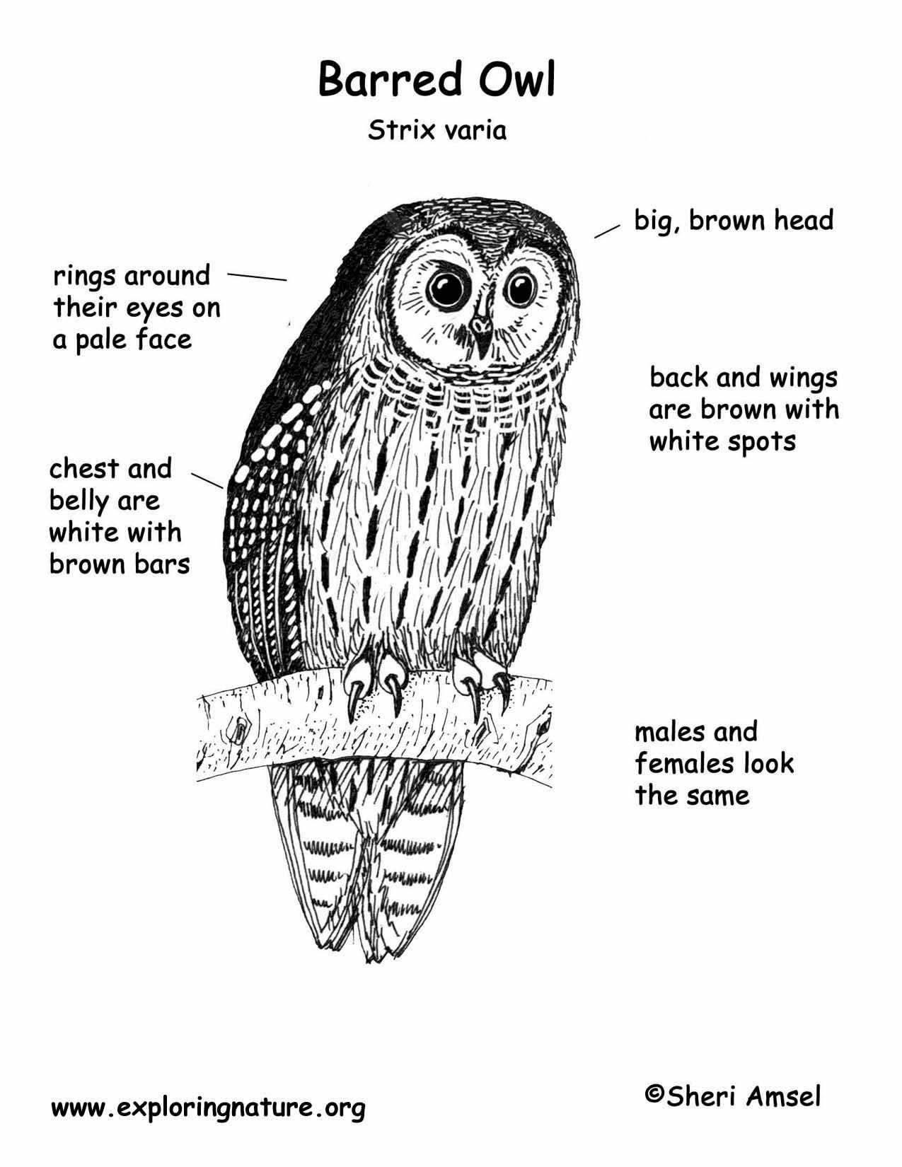
Owl (Barred)
Nucleotide - Genome.gov A nucleotide is the basic building block of nucleic acids (RNA and DNA). A nucleotide consists of a sugar molecule (either ribose in RNA or deoxyribose in DNA) attached to a phosphate group and a nitrogen-containing base. The bases used in DNA are adenine (A), cytosine (C), guanine (G) and thymine (T).
11 Best Images of Blank Anatomy Worksheets - Human Anatomy Body ...
Anatomy And Physiology Archive | June 09, 2022 | Chegg.com Anatomy and physiology archive containing a full list of anatomy and physiology questions and answers from June 09 2022. ... Question 64 Not M T FOR Question Please refer to the following diagram for the next question. 3 K F A Human Nephron In a healthy person, the reabsorption of water back into the bloodstream occurs in w ... 1 answer Eye and ...

Labeled Viking Longship Worksheet - Made By Creative Label
Histology guide: Definition and slides - Kenhub These include epithelial cells, fibroblasts, neutrophils, erythrocytes, keratinocytes, chondrocytes just to name a few. Eukaryotic cell Explore study unit Main tissue types Cells come together with extracellular matrix (a jelly-like fluid) to form the four types of tissues found in the human body: epithelial, connective, muscle and nervous.
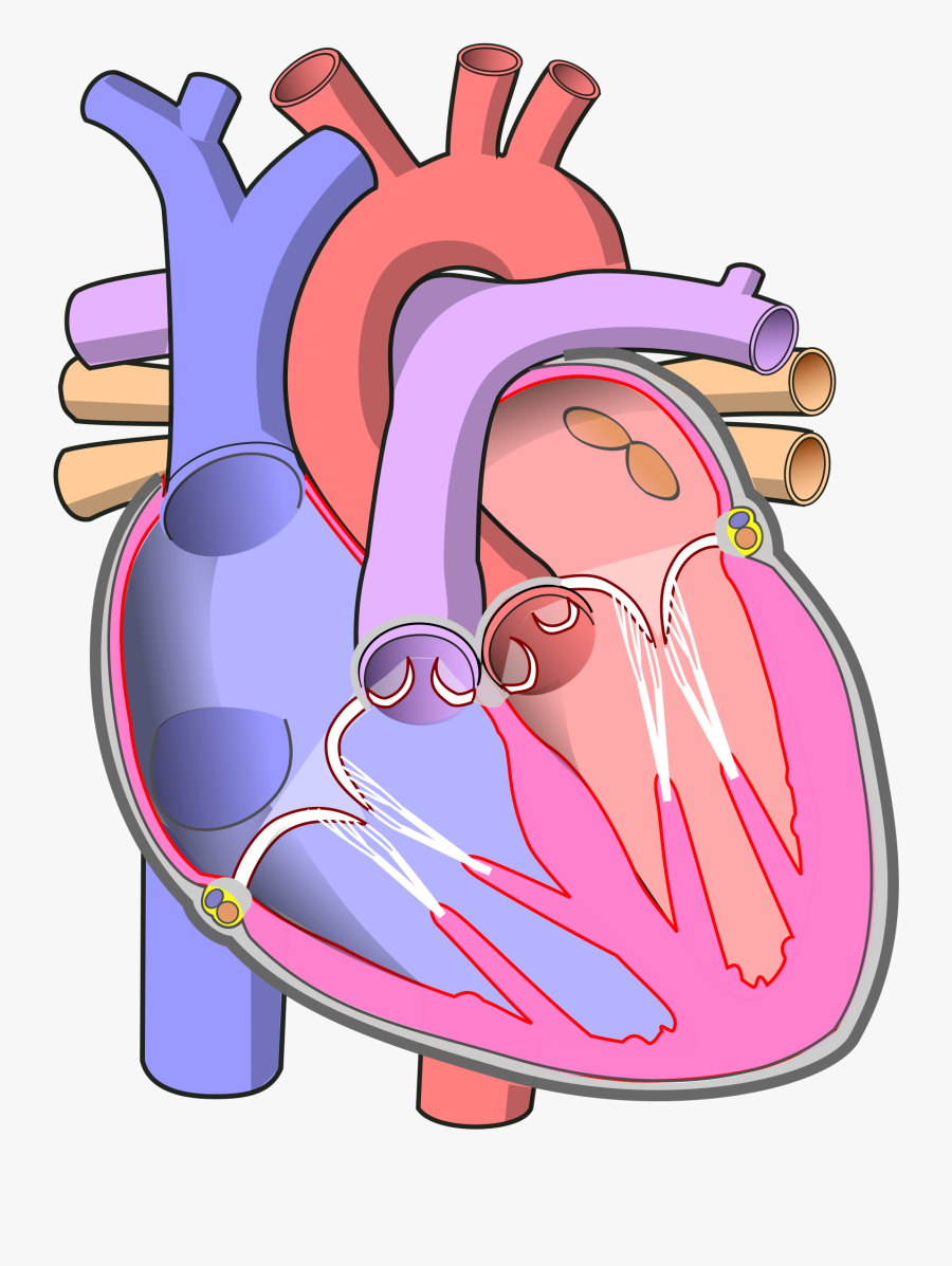
Clip Art File Diagram Of The - Human Heart Without Labels , Free ...
Appendicular Skeleton: Bones List, Diagram & More - Embibe The Human Skeletal system consists of a framework of bones and a few cartilages. This system has a significant role in the movement shown by the body. (Source: NCERT) The skeletal system of humans can be basically divided into two parts (i) the axial skeleton and (ii) the appendicular skeleton.
Post a Comment for "44 simple skeleton diagram to label"