45 labeled eye diagram
Anatomy, Head and Neck, Eye Nerves - StatPearls - NCBI Bookshelf The eyes are a set of sensory organs that play a crucial role in the visual system. The eyes are responsible for detecting light that enters the eyes. Then, the light gets converted into an image in the brain. The sensory and motor innervation of the eyes originate from six paired cranial nerves. These nerves work in sync to manifest movements, reflexes, and vision. Blank ear diagrams and quizzes: The fastest way to learn - Kenhub Take a moment to look at the ear model labeled above. This shows you all of the structures you've just learned about in the video, labeled on one diagram. Seeing them all together in this way is a great way to learn, since anatomical structures do not exist in isolation. That's why labeling the ear is an effective way to begin your revision.
External and internal eye anatomy - MedlinePlus External and internal eye anatomy Overview The cornea allows light to enter the eye. As light passes through the eye the iris changes shape by expanding and letting more light through or constricting and letting less light through to change pupil size. The lens then changes shape to allow the accurate focusing of light on the retina.
Labeled eye diagram
The Best 14 Labeled Anatomy Of Eye Diagram - twomoinfoesz Labeled Anatomy Of Eye Diagram are a subject that is being searched for and appreciated by netizens today. You can Download the Labeled Anatomy Of Eye Diagram here. Download all royalty-free images. Eye Labeling Quizlet - jwc.asl5.piemonte.it The iris lies in front of the crystalline lens and separates the anterior chamber form the posterior chamber Part 3: Labs over the Eye and Ear Label this diagram of the eye (on the ends of the labels) Image: 2bd16821-eaa1-4288-9245-ac87876bf61c Read the definitions below, then label the turkey diagram App Package App Package. Eye Anatomy and Physiology a Complete Detail - Study Read Eyes diagram showing the entire structure The sclera It makes up the outermost part of eye anatomy. It is made of a dense, strong fibrous wall consisting of the sclera that is 5/6 th and the cornea that is anterior 1/6 th of the eyeball. The sclera is the outermost layer and it gives a definite shape to the eye.
Labeled eye diagram. Microscope, Microscope Parts, Labeled Diagram, and Functions 19.01.2022 · Revolving Nosepiece or Turret: Turret is the part of the microscope that holds two or multiple objective lenses and helps to rotate objective lenses and also helps to easily change power. Objective Lenses: Three are 3 or 4 objective lenses on a microscope. The objective lenses almost always consist of 4x, 10x, 40x and 100x powers. The most common eyepiece lens is 10x and when it coupled … Anatomy of the Eye | BrightFocus Foundation Optic nerve: The bundle of nerve fibers at the back of the eye that carry visual messages from the retina to the brain. Photoreceptors: The light sensing nerve cells (rods and cones) located in the retina. Pupil: The adjustable opening at the center of the iris through which light enters the eye. Retina: The light sensitive layer of tissue that ... Duck Anatomy – External and Internal Features with Labeled Diagram 24.07.2021 · Here, I will discuss the body shape, wings, tail, feathers, eye, bill, feet, and more from a duck. ... I hope the duck internal anatomy labeled diagram might help you to identify the structures practically. You might learn the details anatomical facts of bones, muscles, digestive organs from a duck. Categories Avian Anatomy, Veterinary Gross Anatomy Tags avian, duck anatomy, duck anatomy … Parts of Stereo Microscope (Dissecting microscope) – labeled diagram ... Labeled part diagram of a stereo microscope Major structural parts of a stereo microscope. There are three major structural parts of a stereo microscope. The viewing Head includes the upper part of the microscope, which houses the most critical optical components, including the eyepiece, objective lens, and light source of the microscope.
Compound Microscope- Definition, Labeled Diagram, Principle, … 03.04.2022 · The naked eye can now view the specimen at magnification 400 times greater and so microscopic details are revealed. Alternatively, the magnification of the compound microscope is given by: m = D/ f o * L/f e where, D = Least distance of distinct vision (25 cm) L = Length of the microscope tube fo = Focal length of the objective lens fe = Focal length of the eye-piece lens. Parts of a Compound ... Labeled imaging anatomy cases | Radiology Reference Article ... This article lists a series of labeled imaging anatomy cases by body region and modality. Brain CT head: non-contrast axial CT head: non-contrast coronal CT head: non-contrast sagittal CT head: angiogram axial CT head: angiogram coronal CT... Quiz: Label The Parts Of The Eye - ProProfs How much did you get to understand about the human eye? Take up this quiz and find out! Questions and Answers. 1. A is pointing to what part of the eye? A. Cornea. B. Optic Nerve. Eye Anatomy | Blood supply - Orbit - Extraocular muscles | Geeky Medics In total, there are seven extraocular muscles. Six of these are responsible for the movement of the eye, with the seventh being responsible for the movement of the superior eyelid. The muscles responsible for the movement of the eye may be divided into the four recti muscles and the two oblique muscles. Table 1.
What are the 12 cranial nerves? Functions and diagram The ophthalmic part gives sensation to parts of the eyes, including the cornea, mucosa in the nose, and skin on the nose, the eyelid, and the forehead. The maxillary part gives sensation to the... Orbits and eyes: anatomical illustrations - e-Anatomy - IMAIOS Anatomy of the eye : illustrations. Common tendinous ring; Common anular tendon (Zinn) : Extraocular muscles; Extrinsic muscles of eyeball. Superior orbital fissure/Inferior orbital fissure: Nerves, Arteries, Veins. Fascial sheath of eyeball (Tenon) : Extraocular muscles; Extrinsic muscles of eyeball. Eye Diagram Quiz - ProProfs Quiz Try this amazing Eye Diagram Quiz quiz which has been attempted 4816 times by avid quiz takers. Also explore over 77 similar quizzes in this category. Take Quizzes. Animal; Nutrition; ... Quiz: Label The Parts Of The Eye. People say that the eyes are the windows to a person's soul. In the class today, we covered parts of the eye, and what ... Lacrimal apparatus: Anatomy, parts & function | Kenhub The palpebral part lines the internal surface of the eyelids themselves. The bulbar section covers the eyeball. The fornix section is the flexible region that connects the tarsal and bulbar parts, and allows the eye to move with freedom. Bulbar conjunctiva (cranial view)
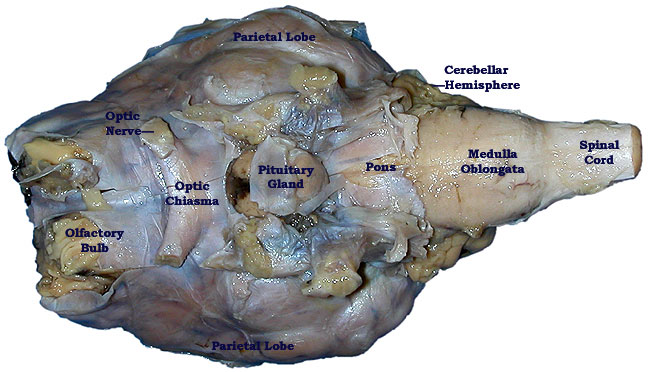
Sheep Brain Dissection Bi - BIOLOGY JUNCTION
Male Human Anatomy Diagram Pictures, Images and Stock Photos Man diagram x-ray cardiovascular, nervous, limphatic and skeletal systems. Man diagram x-ray cardiovascular, nervous, limphatic and skeletal systems. On black background. male human anatomy diagram stock pictures, royalty-free photos & images

Sea Turtle Physical Examination Part 1: Eyes-Ears-Nose-Throat | LafeberVet
Anatomy of the eye: Quizzes and diagrams | Kenhub One of our favorite ways to get to grips with all of the parts of the eye is by utilizing labeled diagrams. On a diagram of the eye, we can see all of the relevant structures together on one image. This helps us to understand how each one is situated and related to the other. Labeled diagram of the eye
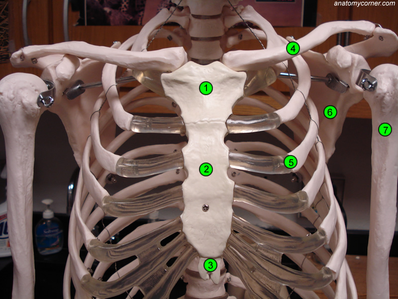
chest.jpg
Illustrations and diagrams of the 12 pairs of cranial nerves - e-Anatomy This human anatomy module is about the cranial nerves. It consists of 15 vector anatomical drawings with 280 anatomical structures labeled. It is intended for the use of medical students working on human anatomy, student nurses, physiotherapists, electro-radiological technicians and residents - especially those working in neurology, neurosurgery, otolaryngology - and for any physician ...

Pin by Laura Hutchinson on Anatomy | Eye anatomy, Anatomy, Anatomy ...
[Human Eye Anatomy And Physiology] - 16 images - human anatomy, anatomy ... [Human Eye Anatomy And Physiology] - 16 images - bones skull frontal anatomy physiology, cow eye dissection diagram labeled featuresofthe cat eye internal, back pain gifs on giphy, eye anatomy and physiology nurseinfo,

eye diagram - PurposeGames
Orbital septum | Radiology Reference Article | Radiopaedia.org The orbital septum (plural: orbital septa) is a thin sheet of fibrous tissue that originates from the orbital rim periosteum and blends with the tendon of the levator palpebrae superioris superiorly and inserts into the tarsal plate inferiorly. The orbital septum separates the intra-orbital fat from eyelid fat and orbicularis oculi muscle, and ...
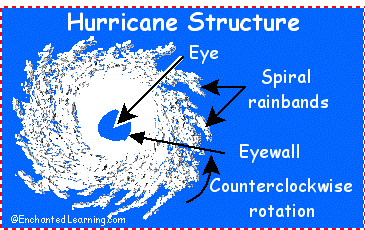
Hurricane Structure: EnchantedLearning.com
A List of Bones in the Human Body With Labeled Diagrams These are bones of the toes of the feet. There are 5 proximal phalanges in each foot (as shown in the diagram above). There are 4 intermediate phalanges, one on each finger, except the big toe. The phalanges on the tips of the toes are known as the distal phalanges. They are 5 in number. In addition to the bones mentioned above, there is ...

Labeling Parts Of the Heart Lovely Parts the Heart Proprofs Quiz ...
Parts of a microscope with functions and labeled diagram 19.04.2022 · Q. Differentiate between a condenser and an Abbe condenser. Ans. Condensers are lenses that are used to collect and focus light from the illuminator into the specimen. They are found under the stage next to the diaphragm of the microscope. They play a major role in ensuring clear sharp images are produced with a high magnification of 400X and above.

Label the Eye
Eye Pressure - American Academy of Ophthalmology Eye pressure—also called intraocular pressure or IOP—is a measurement of the fluid pressure inside the eye. Measuring it is like measuring blood pressure.. The eye has a jelly-like substance called vitreous humor filling most of the back part of the eye. A more-watery liquid called aqueous humor also is present. Much of the aqueous humor is in the front part of the eye, behind the cornea ...

Eye Diagram Without Labels | via Anatomy Pictures Gallery if… | Flickr
Parts of Human Eye and Their Functions - MD-Health.com The iris is the area of the eye that contains the pigment which gives the eye its color. This area surrounds the pupil, and uses the dilator pupillae muscles to widen or close the pupil. This allows the eye to take in more or less light depending on how bright it is around you. If it is too bright, the iris will shrink the pupil so that they ...
Post a Comment for "45 labeled eye diagram"