38 image of microscope with label
Microscope Labeled Pictures, Images and Stock Photos Microscope Labeled Pictures, Images and Stock Photos View microscope labeled videos Browse 49 microscope labeled stock photos and images available, or start a new search to explore more stock photos and images. Newest results Fluorescent Imaging immunofluorescence of cancer cells growing... Microscope diagram vector illustration. Microscope Parts, Function, & Labeled Diagram - slidingmotion Microscope parts labeled diagram gives us all the information about its parts and their position in the microscope. Microscope Parts Labeled Diagram The principle of the Microscope gives you an exact reason to use it. It works on the 3 principles. Magnification Resolving Power Numerical Aperture. Parts of Microscope Head Base Arm Eyepiece Lens
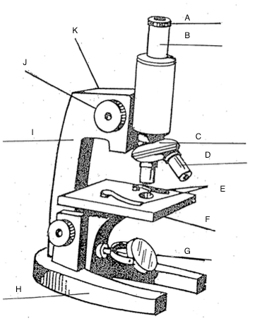
Microscope Coloring - The Biology Corner Light microscopes use either a bulb or a mirror (M) as their light source. Color the light source yellow . The switch for this light is usually found on the base of the microscope, and sometimes on the power cord. You can control how much light goes through the specimen by adjusting the diaphragm (K). Color the diaphragm light green .

Image of microscope with label
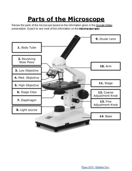
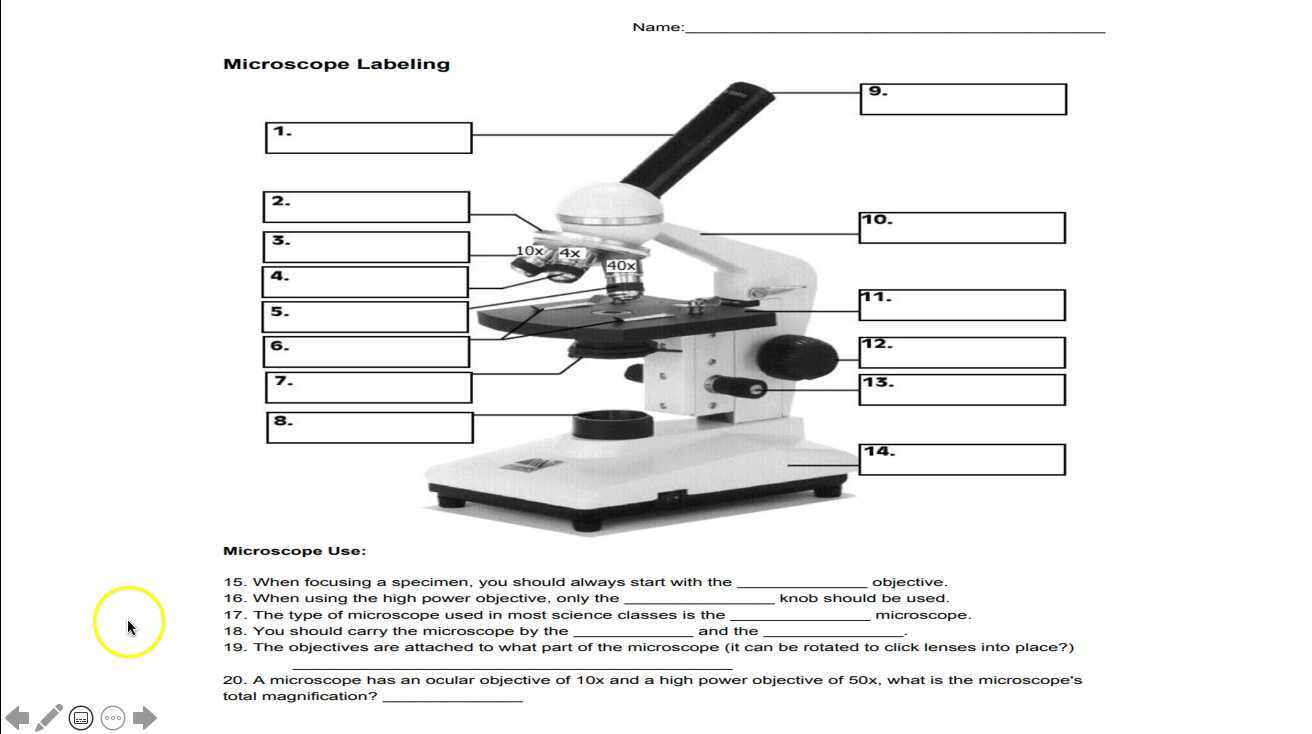
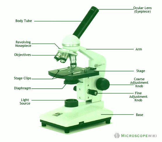
Microscope Labeling - The Biology Corner The labeling worksheet could be used as a quiz or as part of direct instruction where students label the microscope as you go over what each part is used for. The google slides shown below have the same microscope image with the labels for students to copy. I often spend the first day walking students through the steps and having them look at a ... Microscope, Microscope Parts, Labeled Diagram, and Functions Revolving Nosepiece or Turret: Turret is the part of the microscope that holds two or multiple objective lenses and helps to rotate objective lenses and also helps to easily change power. Objective Lenses: Three are 3 or 4 objective lenses on a microscope. The objective lenses almost always consist of 4x, 10x, 40x and 100x powers. The most common eyepiece lens is 10x and when it coupled with ... Labelled Diagram of Compound Microscope The below mentioned article provides a labelled diagram of compound microscope. Part # 1. The Stand: The stand is made up of a heavy foot which carries a curved inclinable limb or arm bearing the body tube. The foot is generally horse shoe-shaped structure (Fig. 2) which rests on table top or any other surface on which the microscope in kept.
Image of microscope with label. Parts of a Simple Microscope - Labeled (with diagrams) A simple microscope is a very first type of microscope ever created. It consists of simple parts and performs simple functions. Although there are now many advanced microscope types, some applications may still demand the use of a simple microscope. In this article, we are going to discuss the parts and functions of a simple microscope. en.wikipedia.org › wiki › Microscope_slideMicroscope slide - Wikipedia A microscope slide is a thin flat piece of glass, typically 75 by 26 mm (3 by 1 inches) and about 1 mm thick, used to hold objects for examination under a microscope. Typically the object is mounted (secured) on the slide, and then both are inserted together in the microscope for viewing. This arrangement allows several slide-mounted objects to ... Compound Microscope Parts - Labeled Diagram and their Functions The eyepiece (or ocular lens) is the lens part at the top of a microscope that the viewer looks through. The standard eyepiece has a magnification of 10x. You may exchange with an optional eyepiece ranging from 5x - 30x. [In this figure] The structure inside an eyepiece. The current design of the eyepiece is no longer a single convex lens. Microscope Parts and Functions Most specimens are mounted on slides, flat rectangles of thin glass. The specimen is placed on the glass and a cover slip is placed over the specimen. This allows the slide to be easily inserted or removed from the microscope. It also allows the specimen to be labeled, transported, and stored without damage.
Compound Microscope Parts, Functions, and Labeled Diagram Compound Microscope Definitions for Labels. Eyepiece (ocular lens) with or without Pointer: The part that is looked through at the top of the compound microscope. Eyepieces typically have a magnification between 5x & 30x. Monocular or Binocular Head: Structural support that holds & connects the eyepieces to the objective lenses. Compound Microscope - Diagram (Parts labelled), Principle and Uses See: Labeled Diagram showing differences between compound and simple microscope parts Structural Components The three structural components include 1. Head This is the upper part of the microscope that houses the optical parts 2. Arm This part connects the head with the base and provides stability to the microscope. Microscope picture label Flashcards | Quizlet Start studying Microscope picture label. Learn vocabulary, terms, and more with flashcards, games, and other study tools. › en › microscopeMicroscope Image Analysis Software | OLYMPUS Stream | Olympus For advanced micro-imaging software that allows you to seamlessly acquire, process, and measure images, our Olympus Stream is the perfect model for you.
Microscope Labeling - The Biology Corner Students label the parts of the microscope in this photo of a basic laboratory light microscope. Can be used for practice or as a quiz. ... Microscope Labeling . Microscope Use: 15. When focusing a specimen, you should always start with the _____ objective. 16. When using the high power objective, only the _____ knob should be used. 17. The ... www1.udel.edu › biology › ketchamMicroscopy Pre-lab Activities - University of Delaware Microscope controls: turn knobs (click and hold on upper or lower portion of knob) throw switches (click and drag) turn dials (click and drag) move levers (click and drag) changes lenses (click and drag on objective housing) select a specimen (click on a slide) 300+ Free Microscope & Laboratory Images - Pixabay 399 Free images of Microscope Related Images: laboratory science bacteria research scientist lab biology chemistry medical Find your perfect microscope image. Free pictures to download and use in your next project. rsscience.com › stereo-microscopeParts of Stereo Microscope (Dissecting microscope) – labeled ... Unlike a compound microscope that offers a flat image, stereo microscopes give the viewer a 3-dimensional image that you can see the texture of a larger specimen. [In this image] Examples of Stereo & Dissecting microscopes. Major microscope brands (Zeiss, Olympus, Nikon, Amscope, Omano, Leica …) all produce stereomicroscopes.
Compound Microscope Parts, Diagram Definition, Application ...
465,820 Microscope Images, Stock Photos & Vectors | Shutterstock Microscope royalty-free images 465,820 microscope stock photos, vectors, and illustrations are available royalty-free. See microscope stock video clips Image type Orientation Color People Artists More Sort by Popular Science College and University Healthcare and Medical Jobs/Professions Biology microscope laboratory scientist medicine lens Next

Microscope Diagram and Functions | Microscope parts, Science ...
Simple Microscope - Diagram (Parts labelled), Principle, Formula and Uses The working principle of a simple microscope is that when a lens is held close to the eye, a virtual, magnified and erect image of a specimen is formed at the least possible distance from which a human eye can discern objects clearly. Magnification formula The magnification power of a simple microscope is expressed as: M = 1 + D/F Where

Biology label part of microscope
Label the microscope — Science Learning Hub All microscopes share features in common. In this interactive, you can label the different parts of a microscope. Use this with the Microscope parts activity to help students identify and label the main parts of a microscope and then describe their functions. Drag and drop the text labels onto the microscope diagram.

Activity 1).docx - Activity #1 THE MICROSCOPE I. A. Label the ...
Microscope Images at Various Magnifications | Microscope World Resources The compound microscope typically has three or four magnifications - 40x, 100x, 400x, and sometimes 1000x. At 40x magnification you will be able to see 5mm. At 100x magnification you will be able to see 2mm. At 400x magnification you will be able to see 0.45mm, or 450 microns. At 1000x magnification you will be able to see 0.180mm, or 180 microns.

Parts of a Microscope - SmartSchool Systems
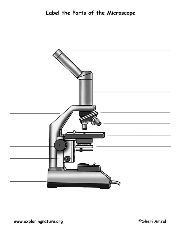
Parts of the Microscope with Labeling (also Free Printouts) Microscopes are specially created to magnify the image of the subject being studied. This exercise is created to be used in homes and schools. the microscope layout, including the blank and answered versions are available as pdf downloads. Click to Download : Label the Parts of the Microscope (A4) PDF print version.

label the parts of the compound microscope - Brainly.ph
en.wikipedia.org › wiki › Microscope_image_processingMicroscope image processing - Wikipedia Microscope image processing is a broad term that covers the use of digital image processing techniques to process, analyze and present images obtained from a microscope. Such processing is now commonplace in a number of diverse fields such as medicine, biological research, cancer research, drug testing, metallurgy, etc. A number of ...
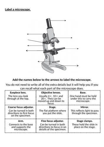
Label a microscope - Teaching resources
Microscope Stock Photos, Pictures & Royalty-Free Images - iStock Microscope Pictures, Images and Stock Photos View microscope videos Browse 196,265 microscope stock photos and images available, or search for magnifying glass or microscope isolated to find more great stock photos and pictures. Newest results magnifying glass microscope isolated science microscope icon laboratory scientist microscope

E-Katalog 5.0
Microscope Labeling Game - PurposeGames.com About this Quiz. This is an online quiz called Microscope Labeling Game. There is a printable worksheet available for download here so you can take the quiz with pen and paper. This quiz has tags. Click on the tags below to find other quizzes on the same subject. Science.

MICROSCOPE Labeling - Part - 3
22 Parts Of a Microscope With Their Function And Labeled Diagram A microscope is a laboratory instrument used to examine objects that are too small to be seen by the naked eye. In other words, it enlarges images of small objects. Invented by a Dutch spectacle maker in the late 16th century, light microscopes use lenses and light to magnify images.
Microscope | Other Quiz - Quizizz
› p-3470-what-is-aWhat is a Compound Microscope? | Microscope World Blog A compound microscope provides a two-dimensional image, while a stereo microscope provides a three-dimensional image. Compound microscopes typically provide magnification in the range of 40x-1000x, while a stereo microscope will provide magnification of 10x-40x.
Parts of the Microscope Labeling Activity!
Microscope Types (with labeled diagrams) and Functions The working principle of a simple microscope is that when a lens is held close to the eye, a virtual, magnified and erect image of a specimen is formed at the least possible distance from which a human eye can discern objects clearly. Simple microscope labeled diagram Simple microscope functions It is used in industrial applications like:

Microscope Labeling Activity - SMART Board Activity - Interactive Review
Simple Microscope - Parts, Functions, Diagram and Labelling What is good about transmission electron microscope is that it provides a high degree of magnification and resolution. It is useful in various fields of sciences such as physical and biological science, nanotechnology, metallurgy, and forensic analysis. (1, 2, 3, and 4) Picture 1: The image above is a stereo microscope.
Microscope Labeling Activity
18,701 Microscope drawing Images, Stock Photos & Vectors - Shutterstock Find Microscope drawing stock images in HD and millions of other royalty-free stock photos, illustrations and vectors in the Shutterstock collection. Thousands of new, high-quality pictures added every day.

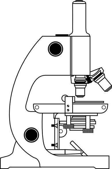
How to Draw a Microscope and Label Its Parts
A Study of the Microscope and its Functions With a Labeled Diagram ... The camera present within the microscope captures images to reveal the finer details of the specimen. This microscope can zoom and view the density of a specimen until it is only a micrometer thick and has a magnification ranging between 1,000 - 250,000x on the fluorescent screen. This microscope needs a computer software to yield precise ...
Mikroskop Binoculer Binokular Microscope Binocular Binokuler XSZ 107 BN XSZ-107BN
Labeling the Parts of the Microscope | Microscope World Resources Labeling the Parts of the Microscope This activity has been designed for use in homes and schools. Each microscope layout (both blank and the version with answers) are available as PDF downloads. You can view a more in-depth review of each part of the microscope here. Download the Label the Parts of the Microscope PDF printable version here.

Compound Microscope Parts – Labeled Diagram and their ...
Parts of a microscope with functions and labeled diagram - Microbe Notes Parts of a microscope with functions and labeled diagram April 19, 2022 by Faith Mokobi Having been constructed in the 16th Century, Microscopes have revolutionalized science with their ability to magnify small objects such as microbial cells, producing images with definitive structures that are identifiable and characterizable.
![750+ Microscope Pictures [HD] | Download Free Images on Unsplash](https://media.istockphoto.com/photos/microscope-isolated-against-white-background-picture-id484035838?b=1&k=20&m=484035838&s=170667a&w=0&h=jNZti7V33guGvJsn5Tq6Dhvr43XOuCpgQ4CIMOf55js=)
750+ Microscope Pictures [HD] | Download Free Images on Unsplash
› techniques › fluorescenceIntroduction to Fluorescence Microscopy | Nikon’s MicroscopyU As presented in Figure 1, the reflected light vertical illuminator comprises an arc-discharge lamphouse at the rear end (usually a mercury or xenon burner).Excitation light travels along the illuminator perpendicular to the optical axis of the microscope, passes through collector lenses and a variable, centerable aperture diaphragm, and then through a variable, centerable field diaphragm (see ...
The Microscope
Labeled Microscope and Basics of Life Diagram | Quizlet PLAY. A microscope is an instrument widely to magnify and resolve the image of an object that is otherwise invisible to naked eye. For resolving the details of objects, which otherwise cannot be achieved by naked eye, a microscope is used. This set of flash cards will help the student to identify the different parts and function of the microscope.

Microscope World Blog: Labeling the Parts of the Microscope
Labelled Diagram of Compound Microscope The below mentioned article provides a labelled diagram of compound microscope. Part # 1. The Stand: The stand is made up of a heavy foot which carries a curved inclinable limb or arm bearing the body tube. The foot is generally horse shoe-shaped structure (Fig. 2) which rests on table top or any other surface on which the microscope in kept.

Label the microscope — Science Learning Hub
Microscope, Microscope Parts, Labeled Diagram, and Functions Revolving Nosepiece or Turret: Turret is the part of the microscope that holds two or multiple objective lenses and helps to rotate objective lenses and also helps to easily change power. Objective Lenses: Three are 3 or 4 objective lenses on a microscope. The objective lenses almost always consist of 4x, 10x, 40x and 100x powers. The most common eyepiece lens is 10x and when it coupled with ...

The Parts of a Microscope (Labeled) Printable Printable (6th ...
Microscope Labeling - The Biology Corner The labeling worksheet could be used as a quiz or as part of direct instruction where students label the microscope as you go over what each part is used for. The google slides shown below have the same microscope image with the labels for students to copy. I often spend the first day walking students through the steps and having them look at a ...
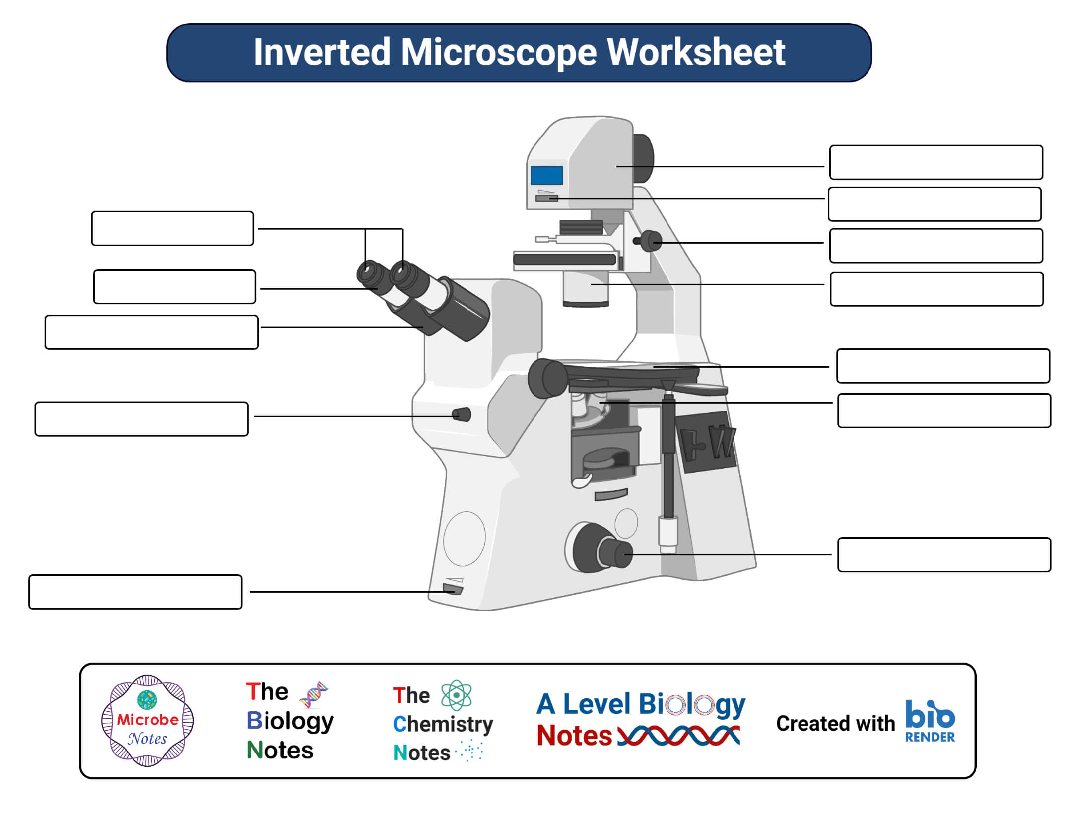
Parts of a microscope with functions and labeled diagram

Microscope Maintenance Tips | Science supplies, Microscope ...

Parts of a microscope with functions and labeled diagram
Lab - Microscope: MAH-Summer 2019-Anatomy and Physiology I
Microscope Labeling
Microscope Types (with labeled diagrams) and Functions
Parts of a microscope with functions and labeled diagram

All About Scopes Labeling A Microscope Ocular Lens

This is a common compound microscope. Label its parts from A ...

Labeling a Microscope Free Worksheet Pack

Produk Microscope | UD Berkah Abadi

A labeled diagram of a microscope. MLT 101. :) | Medical lab ...
Microscope With Labels clip art Vectors graphic art designs ...
7Ac Microscope Labelling Worksheet | Teaching Resources

2.1 " Compound Microscope" | Download Scientific Diagram

Microscope labeled diagram

Compound Microscope Parts – Labeled Diagram and their ...
Post a Comment for "38 image of microscope with label"