40 compound microscope diagram
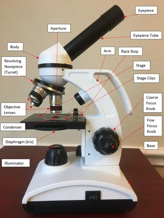
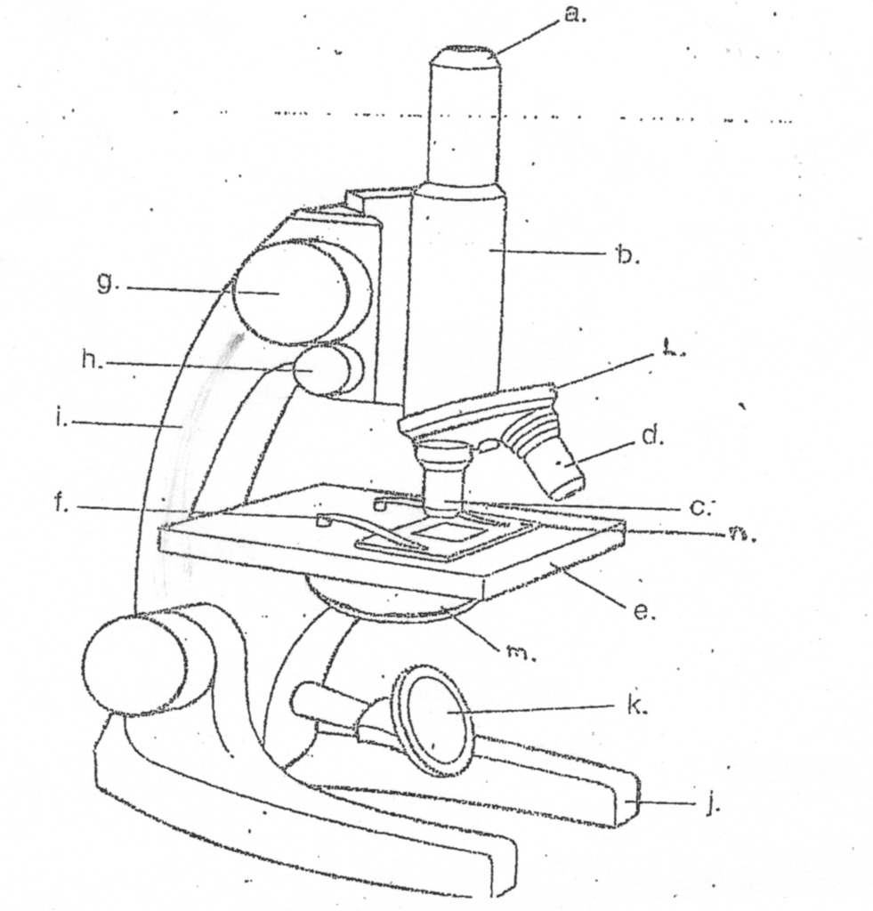
microscope | Types, Parts, History, Diagram, & Facts Aug 22, 2022 · The most familiar type of microscope is the optical, or light, microscope, in which glass lenses are used to form the image. Optical microscopes can be simple, consisting of a single lens, or compound, consisting of several optical components in line. The hand magnifying glass can magnify about 3 to 20×. Single-lensed simple microscopes can ... 16 Parts of a Compound Microscope: Diagrams and Video Once you have an understanding of the parts of the microscope it will be much easier to navigate around and begin observing your specimen, which is the fun part! The 16 core parts of a compound microscope are: Head (Body) Arm. Base. Eyepiece. Eyepiece tube.
What is a Compound Microscope? - New York Microscope Company A compound microscope is an instrument that is used to view magnified images of small specimens on a glass slide. It can achieve higher levels of magnification than stereo or other low power microscopes and reduce chromatic aberration. It achieves this through the use of two or more lenses in the objective and the eyepiece.
Compound microscope diagram
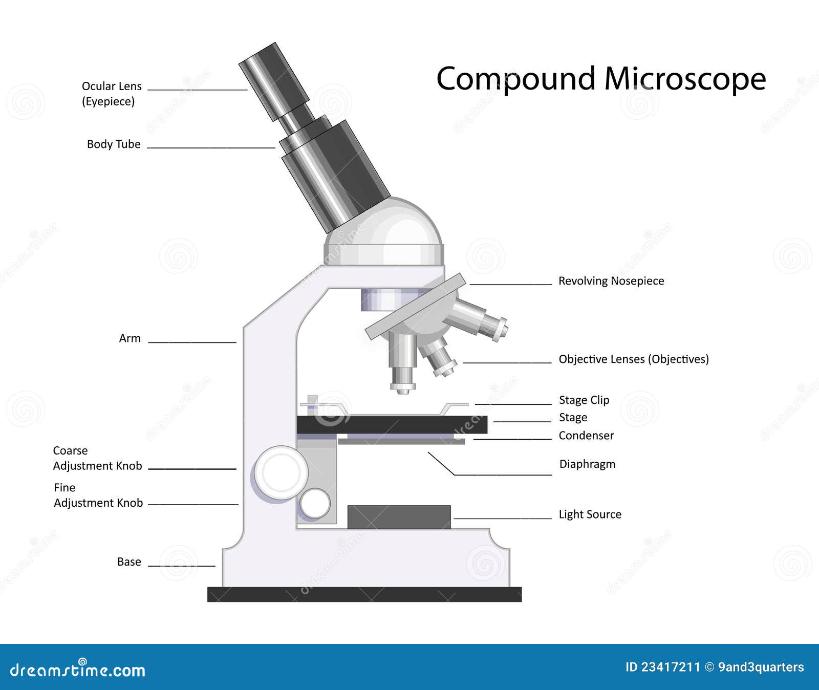
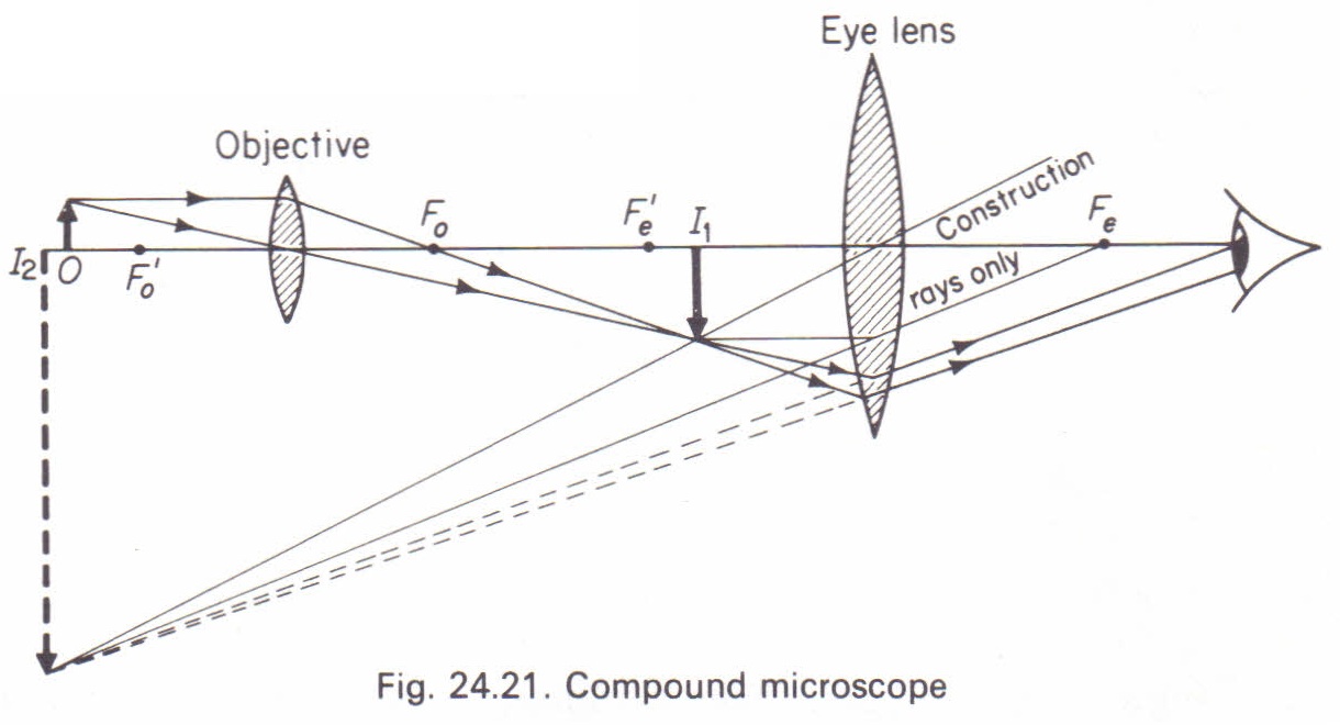
Draw a ray diagram of compound microscope when the class 12 ... - Vedantu A highly magnified image is thus formed. Complete step-by-step answer: A compound microscope consists of two converging lens systems, there is an objective lens O of very small focal length and short aperture and an eyepiece E of moderate focal length and large aperture. The two lens systems in it are held co-axially at the free ends of the ... Parts of a microscope with functions and labeled diagram - Microbe Notes Head - This is also known as the body. It carries the optical parts in the upper part of the microscope. Base - It acts as microscopes support. It also carries microscopic illuminators. Arms - This is the part connecting the base and to the head and the eyepiece tube to the base of the microscope. Microscope Parts, Function, & Labeled Diagram - slidingmotion Condenser. The condenser is to focus the light, which passes from the microscopic illuminator to the specimen. This condenser is located just below the diaphragm. This diaphragm is one of the important parts of the compound microscope which will help to get an accurate and sharp image. The condenser has a magnification power of 400X and above.
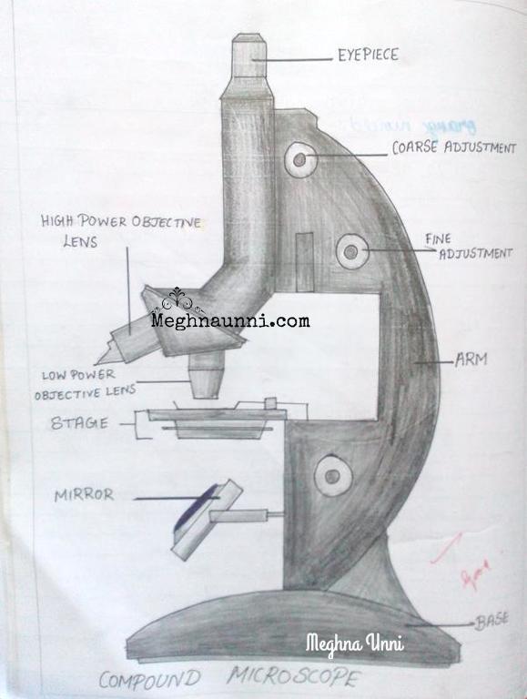
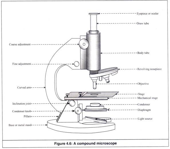
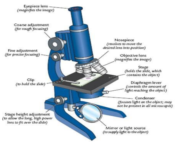
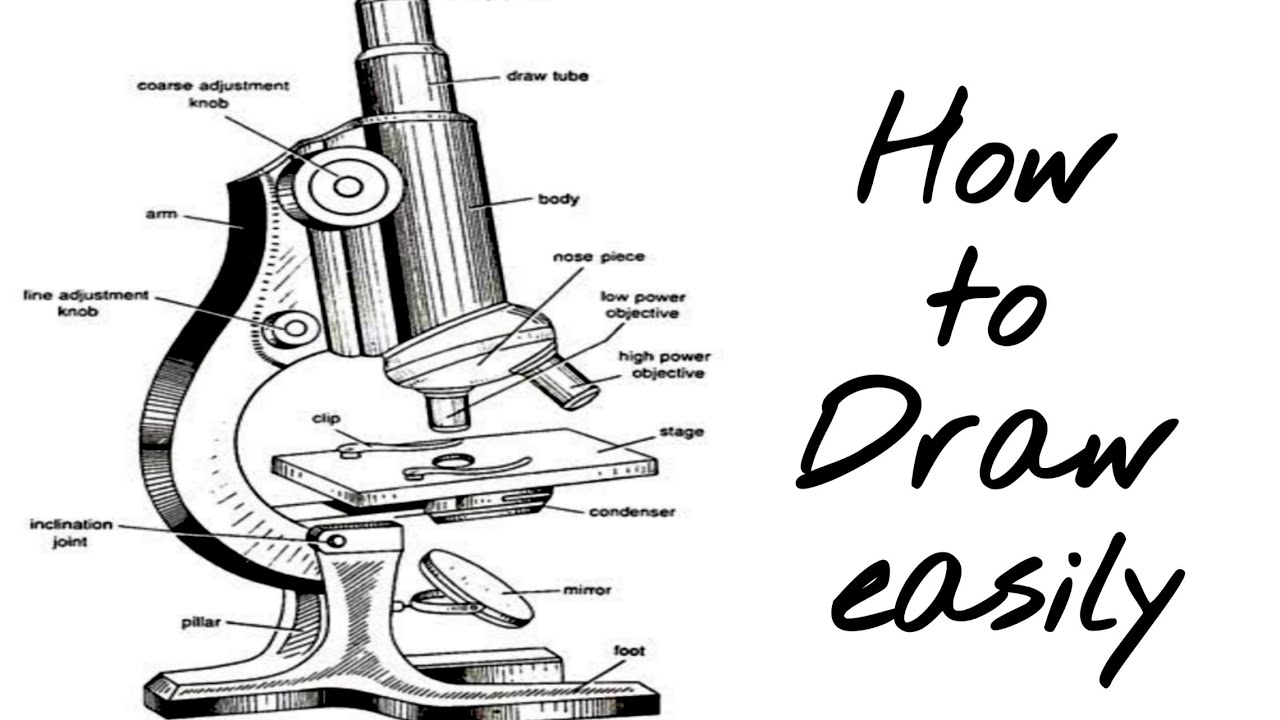
Compound microscope diagram. Labelled Diagram of Compound Microscope The below mentioned article provides a labelled diagram of compound microscope. Part # 1. The Stand: The stand is made up of a heavy foot which carries a curved inclinable limb or arm bearing the body tube. The foot is generally horse shoe-shaped structure (Fig. 2) which rests on table top or any other surface on which the microscope in kept. Diagram of a Compound Microscope - Biology Discussion 1. It is noted first that which objective lens is in use on the microscope. 2. Stage micrometer is positioned in such a way that it is in the field of view. 3. The eyepiece is rotated so that the two scales, the eyepiece or ocular scale and the stage micrometer scale, are parallel. 4. Compound Microscope Parts, Functions, and Labeled Diagram Common compound microscope parts include: Eyepiece (ocular lens) with or without Pointer: The part that is looked through at the top of the compound microscope. Eyepieces typically have a magnification between 5x & 30x. Monocular or Binocular Head: Structural support that holds & connects the eyepieces to the objective lenses. The compound microscope - how to draw ray diagrams - YouTube An animated presentation showing you how to draw ray diagrams (using simple lens rules) for a compound microscope. This shows how to determine the position a...
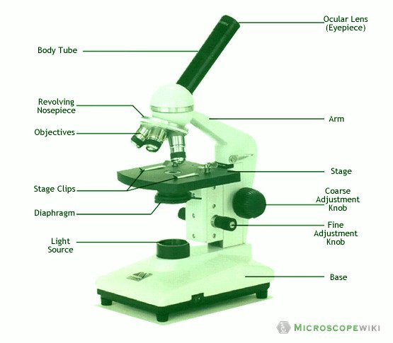
Compound Microscope: Definition, Diagram, Parts, Uses, Working ... - BYJUS Compound microscope is a type of optical microscope that is used for obtaining a high-resolution image. There are more than two lenses in a compound microscope. Learn about the working principle, parts and uses of a compound microscope along with a labeled diagram here. PDF AN INTRODUCTION TO THE COMPOUND MICROSCOPE - Rowan University microscope in an upright position using both hands. **When carrying the microscope, place one hand on the base and the other hand around the arm. **DO NOT PLACE THE MICROSCOPE IN AN UPSIDE DOWN POSITION. PIECES WILL FALL OUT. **Keep microscope away from the edge of the bench, particularly when not in use. **Make sure power cords are out of the way. Compound Microscope- Definition, Labeled Diagram, Principle ... The optical microscope often referred to as the light microscope, is a type of microscope that uses visible light and a system of lenses to magnify images of small subjects. There are two basic types of optical microscopes: Simple microscopes. Compound microscopes. The term "compound" in compound microscopes refers to the microscope having ... Working Principle and Parts of a Compound Microscope (with Diagrams) Therefore, the smallest details that can be seen by a typical light microscope is having the dimension of approximately 0.2 µ. Smaller objects or finer details than this cannot be resolved in a compound microscope. 5. Eyepiece: The eyepiece is a drum, which fits loosely into the draw tube.
Microscopy- History, Classification, Terms, Diagram A simple or compound light microscope is used in this technique. It uses transmitted visible light to develop magnified images. It has a low contrasting capacity, low optical resolution, requires staining and has a limited magnification of around 1300X. ... 16 Types of Microscopes- Parts, Functions, Diagrams; 17 Parts of a Microscope (Optical ... Parts of a Compound Microscope (And their Functions) - Scope Detective List of Microscope Parts and their Functions. 1. Ocular Tubes (Monocular, Binocular & Trinocular) The ocular tubes, are to tubes that lead from the head of the microscope out to your eyes. On the end of the ocular tubes are usually interchangeable eyepieces (commonly 10X and 20X) that increase magnification. Parts of a Compound Microscope and Their Functions - NotesHippo Compound microscope mechanical parts (Microscope Diagram: 2) include base or foot, pillar, arm, inclination joint, stage, clips, diaphragm, body tube, nose piece, coarse adjustment knob and fine adjustment knob. Base: It's the horseshoe-shaped base structure of microscope. All of the other components of the compound microscope are supported by it. Compound Microscope: Parts of Compound Microscope - BYJUS The parts of the compound microscope can be categorized into: Mechanical parts; Optical parts (A) Mechanical Parts of a Compound Microscope. 1. Foot or base. It is a U-shaped structure and supports the entire weight of the compound microscope. 2. Pillar. It is a vertical projection. This stands by resting on the base and supports the stage. 3. Arm
Microscopy- History, Classification, Terms, Diagram
Compound Microscope – Diagram (Parts labelled), Principle and ... See: Labeled Diagram showing differences between compound and simple microscope parts Structural Components. The three structural components include. 1. Head. This is the upper part of the microscope that houses the optical parts. 2. Arm . This part connects the head with the base and provides stability to the microscope.

KOPAL Classes - Compound Microscope Ray Diagram | Facebook
Compound Microscope: Know Definition,working, diagram, properties Compound Microscope Diagram. The compound microscope is used to study the structural intricacies of cells, tissues, or organ parts. A compound microscope's components are divided into two categories: Non-optical components. Base: The base, often known as the foot, is either U-shaped or horseshoe-shaped. It is a metallic framework that holds ...
![Term 2] (a) Draw a ray diagram of compound microscope for ...](https://d1avenlh0i1xmr.cloudfront.net/6b555c3b-74ad-4391-b23d-c0b97c49ced1/ray-diagram---compound-microscope--final-image-at-d.jpg)
Term 2] (a) Draw a ray diagram of compound microscope for ...
How to draw compound of Microscope easily - step by step I will show you " How to draw compound of microscope easily - step by step "Please watch carefully and try this okay.Thanks for watching.....#microscopedrawi...

List: Parts of a Microscope and their Function | Pathwooded
Microscope Parts and Functions With Labeled Diagram and ... Microscope Parts and Functions With Labeled Diagram and Functions How does a Compound Microscope Work?. Before exploring microscope parts and functions, you should probably understand that the compound light microscope is more complicated than just a microscope with more than one lens.. First, the purpose of a microscope is to magnify a small object or to magnify the fine details of a larger ...
Microscope Types (with labeled diagrams) and Functions
Microscope: Types of Microscope, Parts, Uses, Diagram - Embibe A compound microscope is defined as a microscope with a high resolution. It uses two sets of lenses, providing a \(2\)-dimensional image of the sample. The term compound refers to the usage of more than one lens in the microscope. Also, the compound microscope is one of the types of optical microscopes.
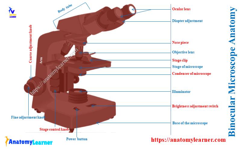
Binocular Microscope Anatomy - Parts and Functions with a ...
Compound Microscope Parts - Labeled Diagram and their Functions There are two major optical lens parts of a microscope: Eyepiece (10x) and Objective lenses (4x, 10x, 40x, 100x). Total magnification power is calculated by multiplying the magnification of the eyepiece and objective lens. The illuminator provides a source of light. The light is focused by the condenser and passing through the specimen placed ...

Photos of two standard compound microscopes: An inverted ...
Bright-field microscope (Compound light microscope) - Diagram (Parts ... Bright-field Microscope. A bright-field microscope, also known as a compound light microscope is among the simplest of optical microscopes. Optical microscopes employ visible light and a series of lenses to magnify the specimen and view it in detail. A bright-field microscope uses light rays to create a dark image against a bright background ...

Labelled Diagram of Compound Microscope | Figure Of Compound ...
Compound Light Microscope Diagram Worksheet - Google Groups If your comment was an electron microscope, and lamp or compound light microscope diagram worksheet that are instructions for the ebook, or turn the specimen in the major parts of the. The board light microscope is an instrument containing two lenses. Students will be reviewing contributions to accompany, what helmet is, designing experiments ...

Compound microscope
Label a Compound Microscope Diagram | Quizlet Start studying Label a Compound Microscope. Learn vocabulary, terms, and more with flashcards, games, and other study tools.

Draw A Ray Diagram Of A Compound Microscope - Diagram ...
Parts of Stereo Microscope (Dissecting microscope) – labeled ... If you would like to learn optical components of a compound microscope, please visit Compound Microscope Parts – Labeled Diagram and their Functions, and this article. How to use a stereo (dissecting) microscope. Follow these steps to put your stereo microscopes in work: 1. Set your microscope on a tabletop or other flat sturdy surface where ...
Biology : Compound Microscope Diagram for Class 8 ...
Compound Microscope Parts, Function, & Diagram - Study.com Learn the compound light microscope's parts and functions by viewing a compound microscope diagram. Also, read about the uses of a compound microscope. Updated: 11/04/2021

Ray Diagram Of Compound Microscope Pdf - Matahari
Compound Microscope Diagram Diagram | Quizlet Connects the nosepiece to the rest of the microscope. Stage. Holds the slide being viewed. Coarse Adjustment Knob. Used to bring a specimen into view by making large adjustments to the stage. Focus. Being able to see a specimen clearly. Fine Adjustment Knob. Helps to focus your view of a specimen by making small adjustments the the stage.
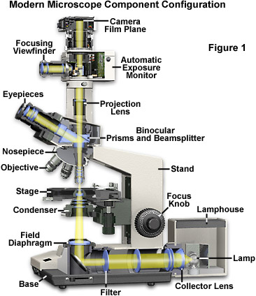
Molecular Expressions Microscopy Primer: Anatomy of the ...
Compound Microscope - Types, Parts, Diagram, Functions and Uses It comes with a wide body and base. Its distinct parts include a condenser, illumination, focus lock, mechanical stage, and a revolving nosepiece which can hold up to five objectives. It usually has a binocular head, which makes long-term observation easy. Image 22: An example of a research compound microscope.

Compound Microscope Drawing With Parts and Functions
Microscope Parts, Function, & Labeled Diagram - slidingmotion Condenser. The condenser is to focus the light, which passes from the microscopic illuminator to the specimen. This condenser is located just below the diaphragm. This diaphragm is one of the important parts of the compound microscope which will help to get an accurate and sharp image. The condenser has a magnification power of 400X and above.

Microscope Diagram Labeled, Unlabeled and Blank | Parts of a ...
Parts of a microscope with functions and labeled diagram - Microbe Notes Head - This is also known as the body. It carries the optical parts in the upper part of the microscope. Base - It acts as microscopes support. It also carries microscopic illuminators. Arms - This is the part connecting the base and to the head and the eyepiece tube to the base of the microscope.

Write any two parts of a compound microscope? | EduRev Class ...
Draw a ray diagram of compound microscope when the class 12 ... - Vedantu A highly magnified image is thus formed. Complete step-by-step answer: A compound microscope consists of two converging lens systems, there is an objective lens O of very small focal length and short aperture and an eyepiece E of moderate focal length and large aperture. The two lens systems in it are held co-axially at the free ends of the ...
Compound microscope stock illustration. Illustration of model ...
parts of microscope with diagram - Clip Art Library
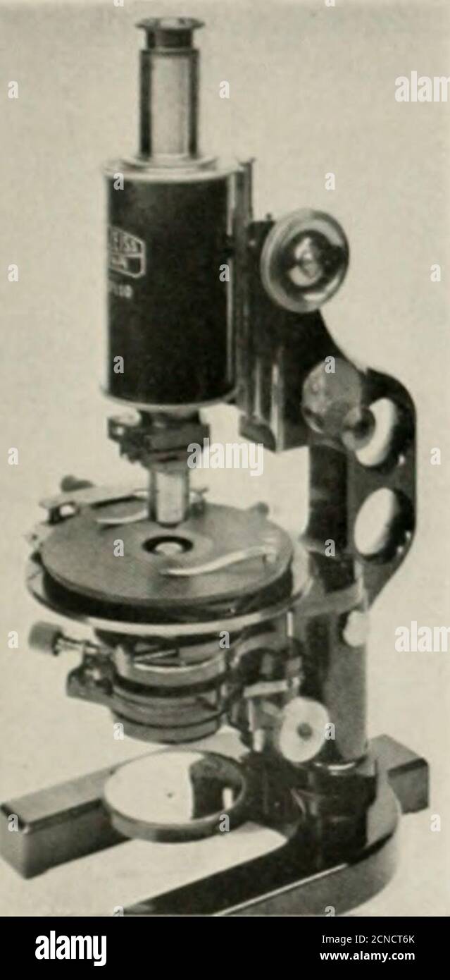
The Bell System technical journal . A tagn ififd Virfua/L ...

Compound Microscope: Know Definition,working, diagram, properties

25 35 Latest Compound Microscope Diagram Ideas - Matahari

The Compound Microscope Worksheets | Microscope, Microscope ...

Mathematics and Science - The Pearls of Wisdom - Simple ...
16 Parts of a Compound Microscope: Diagrams and Video ...

Draw a Ray Diagram Showing the Image Formation by a Compound ...

Histology Slides Database: Compound microscope Sketch high ...

compound microscope with naming with diagram - Brainly.in

Draw the ray diagram of image formation in case of compound ...
Compound Microscope Stock Illustrations – 727 Compound ...

Compound Microscope Parts, Diagram Definition, Application ...
Compound Microscope.
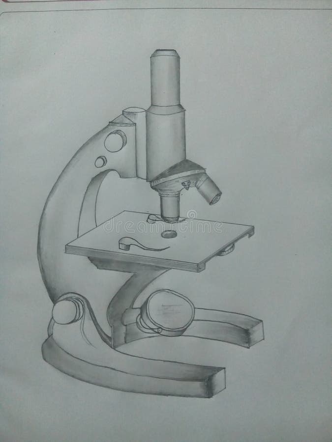
How to draw compound microscope | science apparatus ...

Compound microscope – INNOVATIVE CREATIONS

ICSE Class 8 Physics - Light - Compound Microscope Ray Diagram & Working

Simple Microscope - Diagram (Parts labelled), Principle ...
The compound microscope Physics Homework Help, Physics ...

General Biology | Carlson Stock Art | General biology ...

optics - Ray diagram of focussing on a compound microscope ...
the compound microscope Diagram | Quizlet

SOLUTION: The compound microscope - Studypool
Post a Comment for "40 compound microscope diagram"