42 cow eye parts labeled
Hug machine - Wikipedia A hug machine, also known as a hug box, a squeeze machine, or a squeeze box, is a deep-pressure device designed to calm hypersensitive persons, usually individuals with autism spectrum disorders. The therapeutic, stress-relieving device was invented by Temple Grandin while she was attending college.. Autism has a profound effect upon social interactions and … Parts of the eye, Eye parts, Cow eyes - Pinterest Parts Of An Eye The first line of protection of the eyes is provided by the lids, which prevent access of foreign bodies and assist in the lubrication of the corneal surface. Lid closure and opening are accomplished by the orbicularis oculi and levator palpebri muscles; the orbicularis oculi operates on both lids, bringing their margins into ...
eye anatomy optic nerve Iris eye dissection cow anatomy pupil hole parts main cornea virtual retina. Optic nerve 2. Optic nerve head evaluation in glaucoma eye anatomy optic nerve Cow Eye Dissection we have 7 Pics about Cow Eye Dissection like Human Eye: Anatomy, parts and structure, Unilateral Optic Nerve Hypoplasia in a patient desiring surgical and also Optic nerve 2.

Cow eye parts labeled
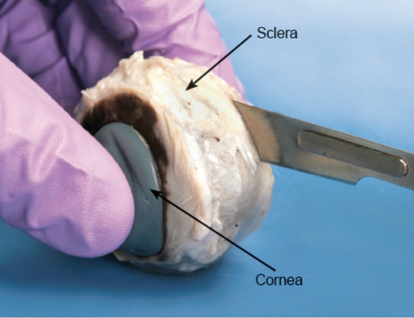
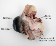
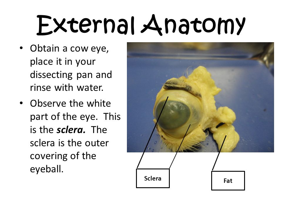
Buying a Cow from a Farmer - Clover Meadows Beef Nov 15, 2021 · Depending on who you order your cow from, you’ll get the option of customizing your cut list. More on that later. At Clover Meadows Beef, if you order a sixteenth, quarter or a half beef, we use a standardized cut list since you’re splitting the cow with other people and the entire cow needs to be processed the same way. If you order a ... Lab 10—Labeled Cow Eye The Cow Eye (Labeled) Return to: Lab 10 PageBIO 137 Main Page Be sure to practice using the unlabeled images. Coronal section, anterior view (lens and vitreous humor displaced) Sagittal section This page created and maintained by Udo M. Savalli. Last updated April 18, 2006. PDF Cow Eye Dissection Lab - Home Science Tools Look carefully at the preserved cow eye. The most noticeable part of the eye is the large mass of gray tissue that surrounds the pos- terior(back) of the eye and is attached to the sclera. The second most noticeable part of the eye is the cornea, located in the ante- rior(front) part of the eye.
Cow eye parts labeled. PDF Cow Eye Dissection: Examining Structure and Function - Woodstown The eyes of cows are structurally and functionally similar to the eyes of humans. During this activity, you will dissect a cow eye. You will observe several important features of the eye and develop your understanding of how each part functions to make vision possible. Materials • Preserved Cow Eye • Scalpel or Scissors • Forceps anatomy and physiology of cow anatomy and physiology of cow anatomy and physiology of cow Sheep brain labeled anatomy external lobe dissection frontal physiology nervous system occipital spinal cord savalli. Cow eye dissection labeled. Sheep heart anatomy and physiology of cow Cow Eye Dissection Teacher's Guide - The Biology Corner I often leave eye models out during the lab to show comparisons between the cow and the human eye. Plus, I've found that my anatomy students have trouble ... National Beef Wire | Cow-Calf Today Home; Channel Guide; Today's Sales
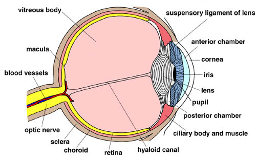
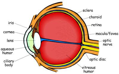
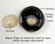
Cow's Eye Dissection - Eye diagram - Exploratorium A cow's pupil is oval. A tough, clear covering over the iris and the pupil that helps protect the eye. Light bends as it passes through the cornea. This is the first step in making an image on the retina. The cornea begins bending light to make an image; the lens finishes the job. The layer of light-sensitive cells at the back of the eye. Cow Eye Dissection & Parts of the Eye Diagram | Quizlet cornea Clear, outer layer of the front of the eye. sclera White, outermost layer of the eye. Helps maintain shape and gives attachment to muscles. photoreceptors The cells in the retina that respond to light (rods and cones) rods Photoreceptor cells in the eye that detect black, white, and gray cones Photoreceptor cells in the eye that detect color Milk - Wikipedia Milk is a nutrient-rich liquid food produced by the mammary glands of mammals.It is the primary source of nutrition for young mammals (including breastfed human infants) before they are able to digest solid food. Immune factors and immune-modulating components in milk contribute to milk immunity.Early-lactation milk, which is called colostrum, contains antibodies that strengthen … Cow Eye for Dissection Specimen - Home Science Tools Cow Eye for Dissection Specimen $3.25 Since a cow's eye is similar to a human's eye, a cow eye dissection is a great way to discover how eyes work! When you order these eyes in bulk, note that you may receive a combo of 10-packs & individually packed specimens. quantity Ages 11+ In Stock & Ready to Ship Need it fast? See delivery options in cart.
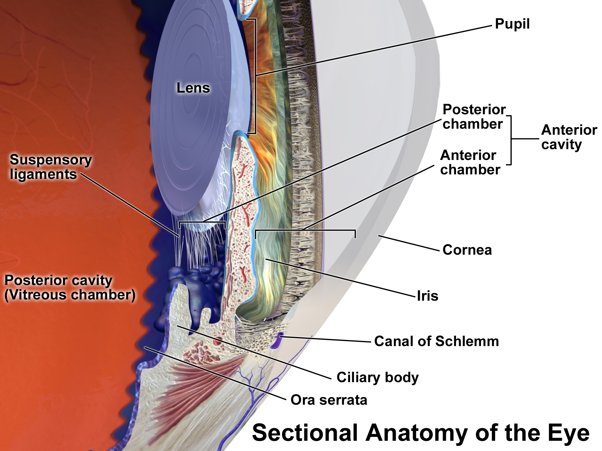
Label Parts of the Human Eye - University of Dayton Parts of the Eye. Select the correct label for each part of the eye. The image is taken from above the left eye. Click on the Score button to see how you did. Incorrect answers will be marked in red. ... PDF Table 8.1: External Anatomy of The Cow Eye Feature Description TABLE 8.1: EXTERNAL ANATOMY OF THE COW EYE FEATURE DESCRIPTION Extrinsic eye muscles Six different muscles attached to the posterior surface of the eyeball that connect it to the bony orbit of the skull Optic nerve Nerve located posteriorly and medially; about 3mm in diameter Conjunctiva Thin, mucous membrane covering the anterior surface of the eyeball; you Is Goat’s Milk Right for You? - Healthline 14.08.2018 · In the case of lactose-free cow’s milk, the lactose is simply split into its component parts (glucose and galactose) so that it’s easier to digest. However, the total sugar count remains constant. Cow Anatomy - External Body Parts and Internal Organs with ... Jul 28, 2021 · In addition, the basal or the ground surface of cow’s hoof consists of two parts – the sole and bulb. The hoof of a cow consists of three parts – peripole, wall, and sole. The peripole surrounds the coronary border, and the wall forms most of the abaxial surface of a hoof. Cow anatomy labeled diagram
Cow Eye Quiz Dissection 101: Click - ppt video online download
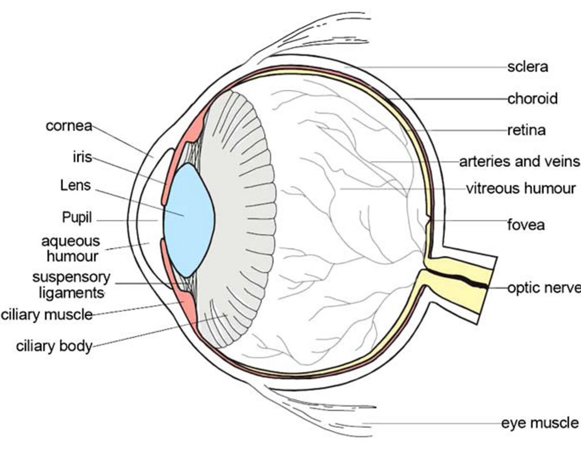
Study 15 Terms | Biology Flashcards | Quizlet 1/15 Created by lilykuchevr Terms in this set (15) Lens 1. Refracts light. 2. Focus light rays on the retina. 3. Located behind the pupil. Retina 1. Sensitive Layer. 2. Detects images. 3. Connects to brain by the optic nerve. 4. Contains cone and cells. Aqueous Humor 1. Watery fluid that keeps the cornea in a rounded shape. Cornea 1. Transparent.
Cow Eye Dissection | Carolina.com
Cow Eye Dissection & Anatomy Project | HST Learning Center Cow Eye Dissection: Internal Anatomy 1. Place the cow's eye on a dissecting tray. The eye most likely has a thick covering of fat and muscle tissue. Carefully cut away the fat and the muscle. As you get closer to the actual eyeball, you may notice muscles that are attached directly to the sclera and along the optic nerve.

Lab#1 Cow Eye Dissection Diagram | Quizlet
Confocal Microscope- Definition, Principle, Parts, Types ... Mar 08, 2022 · In Biomedical sciences, it is used in the analysis of eye corneal infections, by quantifying and qualitatively analyzing the endothelial cells of the cornea. Used to identify the presence of fungal elements in the corneal stroma, during keratomycosis infection, or rapid diagnosis and quick therapeutic response.

Diagram of human eye anatomy with label 1610223 Vector Art at ...
Milk - Wikipedia Cow's milk contains, on average, 3.4% protein, 3.6% fat, and 4.6% lactose, 0.7% minerals and supplies 66 kcal of energy per 100 grams. See also Nutritional value further on in this article and more complete lists at online sources that list values and differences in categories.
Posterior chamber of eyeball - Wikipedia
National Beef Wire | Cattle Network The latest Cattle industry information source on the web. The best cattle and livestock market information at your fingertips.
Cow Eye Dissection: Examining Structure and Function
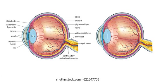
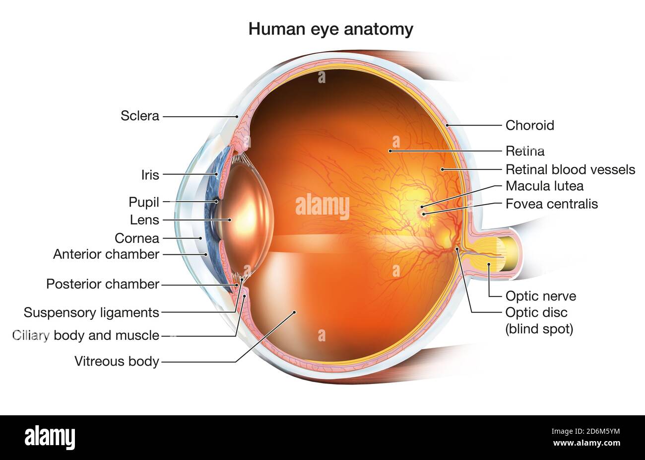
Solved Art-labeling Activity: External and Internal Anatomy - Chegg Expert Answer. 100% (9 ratings) Transcribed image text: Art-labeling Activity: External and Internal Anatomy of the Cow Eye Part A Drag the labels to the appropriate location in the figure. Reset Help Extrinsic muscles of the eye Retina Optic disc (blind spot) Lens Cornea Iris Ciliary body Sclera Optic nerve (cranial nerve II)

Detailed Cow Eye Dissection: Part II (Jr. High, High School and College Review)
National Beef Wire | Cow-Calf Today Home; Channel Guide; Today's Sales
Anatomy of the eye | Children's Wisconsin
PDF Dissection 101: Cow Eye (optic nerve, iris, pupil, sclera, cones, rods, cornea, retina, lens and vitreous humor) Use a labeled drawing if it is helpful. Accept reasonable answers; below is an example of a response for light movement through the eye. Light enters the eye through the transparent cornea which is continuous with the sclera.
NEUR 320: Art and Vision
cow brain anatomy - Microsoft cow eye dissection labeled parts anatomy eyes labels label section diagram structures virtual physiology lab human resize science body tapetum. Brain Of A Cow Stock Photo. Image Of Activity, Abstract - 45875504 . brain cow background. A Day In The Life Of A Cow Vet: She's Crazy!!!

10 Cow eye ideas | cow eyes, anatomy and physiology, eye anatomy
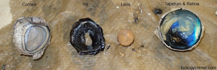
Parts and Functions - Dissecting A COW EYE The four muscles that moves the cow's eye in four directions. Cornea A covering over the iris that helps to protect the eye by making light bend to project the image. Iris and Pupil The iris is a muscle that controls how much light goes into the eye and suspended between the cornea and lens. As we have seen in the dissection a cow's iris is brown.
Cow Eye Dissection | Carolina.com
Duck Anatomy – External and Internal Features with Labeled Diagram 24.07.2021 · After reading this part of the article, you will get a basic idea of the different body parts of a duck. You need to know the following parts from the body of a duck. Bill with bean of a duck; Nostril of a duck; Chin and throat of a duck Crown of a duck; Eye, ear, and nape of a duck; Neck, flank, and abdomen of a duck; Mantle, back, and wing of ...
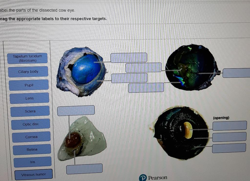
abel the parts of the dissected cow eye. rag the | Chegg.com
Duck Anatomy - External and Internal Features with Labeled ... Jul 24, 2021 · Some other interesting facts about duck anatomy are shown on the labeled diagram at the end of this article. External body parts of duck anatomy. Knowing the external body parts of duck anatomy is very important both for veterinary students and farm owners. Here, I will discuss the body shape, wings, tail, feathers, eye, bill, feet, and more ...
Cow Eye Dissection & Anatomy Project | HST Learning Center
Parts And Functions of The Cow Eye And Conclusion - Weebly COW EYE PARTS: Cornea: A tough clear covering over the iris and pupil that helps protect the eye. ... In conclusion me and Carlo learned a lot about the anatomy of a cow's eyeball. I especially was fascinated by the different parts and functions inside the eyeball. Also dissecting the eyeball was loads of educational entertainment for both of ...
How Vision Works: Eye Science Projects & Experiments | HST
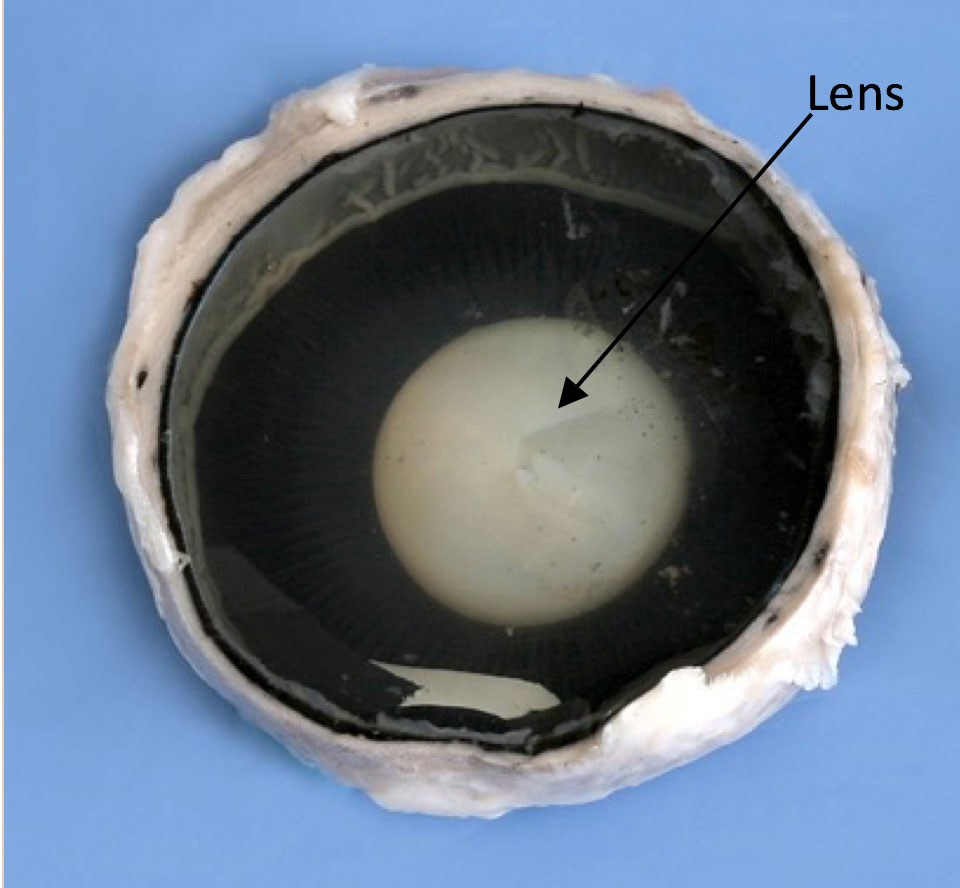
Cow Eye Dissection Guide - Google Slides Cow Eye. Use the point of a scissors or a scalpel to make an incision through the layers of the eye capsule (similar to figure 1); there are three layers from the exterior: sclera, whitish/grey, continuous with the transparent cornea, choroid, thin dark black layer and the retina, thin greyish/pink layer. Use a scissors to dissect the entire ...

Cow's Eye Diagram Quiz
National Beef Wire | Cattle Network The latest Cattle industry information source on the web. The best cattle and livestock market information at your fingertips.
6,847 Human eye diagram Images, Stock Photos & Vectors ...
Mucosa Associated Lymphoid Tissues (MALT) - Microbe Notes 26.06.2018 · Small concentrations of lymphoid tissue are also found in thyroid, breast, lung, salivary glands, eye, and skin. Image Source: DOI: 10.1183/13993003.01701-2015 These lymphoid tissues collectively are thus referred to as …
Cow Eye Dissection
Cow Eye Dissection - The Biology Corner 1. Examine the outside of the eye. You should be able to find the sclera, or the whites of the eye. This tough, outer covering of the eyeball has fat and muscle attached to it 2. Locate the covering over the front of the eye, the cornea. When the cow was alive, the cornea was clear. In your cow's eye, the cornea may be cloudy or blue in color. 2.

Cow Eye Dissection | Carolina.com
label diagram of cow Cow Eye Parts Labeled - All About Cow Photos retina pupil cornea nih keratopathy surgitel buta nampak loupes wagr nerve healthlink sclera Cow Labeling Page labeling Label Turkey Parts Worksheet - Turtle Diary turtlediary

10 Cow eye ideas | cow eyes, anatomy and physiology, eye anatomy
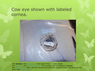
Cow eye - dissection and label - SlideShare Cow eye shown with labeled cornea. The cornea is the transparent front part of the eye that covers the iris, pupil, and anterior chamber. The cornea, with the anterior chamber and lens, refracts light, with the cornea accounting for approximately two-thirds of the eye's total optical power. 3.
Sight — Science Learning Hub
Buying a Cow from a Farmer - Clover Meadows Beef 15.11.2021 · Are you thinking of buying a cow directly ... t pass inspection, it is removed entirely from the food supply. When beef does pass inspection, it is stamped or labeled with the USDA inspection ... For example, at the grocery store you’ll see “round” sold as ground beef, round steak, eye of round, tip steak, tip roast ...

Cow eye – dissection and label
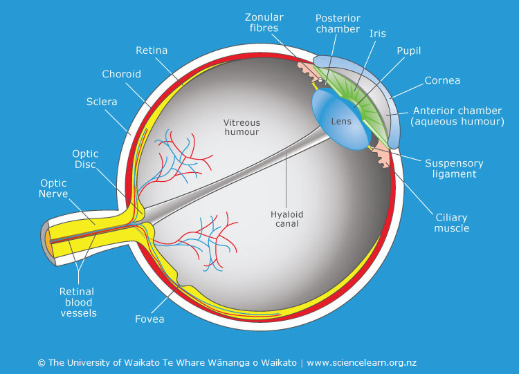
PDF Name: Dissection 101: Cow Eye human eye _____ _____ Draw and label the cow eye. Cornea Optic nerve Vitreous humor Retina Optic disc (blind spot) Choroid Tapetum lucidum Sclera Aqueous humor Suspensory ligaments Lens Ciliary body Pupil Iris Provided by Dissection 101: Cow Eye

The Connection - Join us for a LIVE cow eye dissection with ...
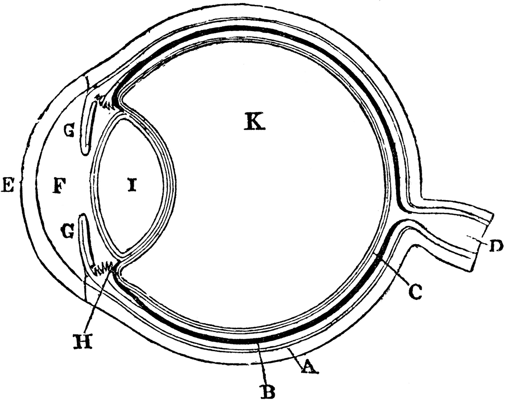
PDF COW'S EYE dissection - Exploratorium This diagram shows the parts of the eye. Can you find these parts in a cow's eye? SCLERA TAPETUM OPTIC NERVE BLIND SPOT LENS VITREOUS HUMOR IRIS CORNEA RETINA AQUEOUS HUMOR PUPIL. COW'S EYE dissection page 3 Examine the outside of the eye. See how many parts of the eye you can identify. You should be able to find

A&P Lab -- Unit 2 -- Labeled Cow Eye Diagram | Quizlet
Confocal Microscope- Definition, Principle, Parts, Types, Labeled ... 08.03.2022 · Types of Confocal Microscope. Confocal laser scanning Microscope – It uses several mirrors that scan along the X and Y axes on the specimen, by scanning and descanning, and the image passes through a pinhole into the detector. Spinning disk, also known as the Nipkow disk, is a type of confocal microscope that uses several movable apertures (pinholes) …
Development, Anatomy and Physiology of the Eye The word ...
Cow Anatomy - External Body Parts and Internal Organs with Labeled … 28.07.2021 · The external body parts from the head region of a cow – in this head region, you might identify the mouth, lip, cheek, chin, muzzle, forehead, poll, ear, eye, nostril, and other. Different parts from the neck region of a cow – here, you will find the neck crest, dewlap, brisket, and jugular groove.

Cow's Eye Dissection - Eye diagram
anatomy of the eye labeled eye cow anatomy diagram labeled dissection eyeball eyes guide lucidum Super Eye Care Resources drshawncohen.com eye anatomy care basics resources 4: Head, Neck, And Dental Anatomy | Pocket Dentistry pocketdentistry.com mandible anatomy dental head neck skull figure pocketdentistry EYE ANATOMY ILLUSTRATION Stock Photo - Alamy

Cow Eye Dissection & Labeling
Cow Eye Dissection | Carolina.com Cow Eye Dissection. The mammalian eye is a sensory organ that operates as part of the nervous system. These complex organs gather light, focus it on receptor cells, and transmit the information to the brain where it is interpreted. Placement and shape of eyes vary across the animal kingdom, but the main function remains consistent—vision.

Eye Diagram Stock Illustrations – 7,502 Eye Diagram Stock ...
PDF Cow Eye Dissection Guide - Central Bucks School District DISSECTION OF THE COW EYE Please make sure to wear gloves and safety glasses when you are dissecting, and make sure to clean up thoroughly after the lab. Also, the cow eyes can be rather slippery, so use caution when handling and cutting them. You will need a scalpel and forceps. 1. First, identify the most external structures of the eye.

Lens (anatomy) - Wikipedia
Parts of a Cow - English Grammar Notes Name of Parts of a Cow Ear Horn Eye Nostril Dewlap Knee Hoof Dewclaw Elbow Udder Teat Toes Hock Stifle Tail Wither Description of the Parts of a Cow on the list Ear The ears of a cow are a sensitive part of their body. The cows use their ears to express their feelings. The ears normally have four different postures.
Cow Eye Dissection: Examining Structure and Function
PDF Cow Eye Dissection Lab - Home Science Tools Look carefully at the preserved cow eye. The most noticeable part of the eye is the large mass of gray tissue that surrounds the pos- terior(back) of the eye and is attached to the sclera. The second most noticeable part of the eye is the cornea, located in the ante- rior(front) part of the eye.

LP 1: Anatomy of cow eye 23.5 Diagram | Quizlet
Lab 10—Labeled Cow Eye The Cow Eye (Labeled) Return to: Lab 10 PageBIO 137 Main Page Be sure to practice using the unlabeled images. Coronal section, anterior view (lens and vitreous humor displaced) Sagittal section This page created and maintained by Udo M. Savalli. Last updated April 18, 2006.
Ciliary body hi-res stock photography and images - Alamy
Buying a Cow from a Farmer - Clover Meadows Beef Nov 15, 2021 · Depending on who you order your cow from, you’ll get the option of customizing your cut list. More on that later. At Clover Meadows Beef, if you order a sixteenth, quarter or a half beef, we use a standardized cut list since you’re splitting the cow with other people and the entire cow needs to be processed the same way. If you order a ...
Using Genetic Algorithm for Identification of Diabetic ...
Cow Eye Labeled Diagram - ClipArt Best

Cow Eye Dissection Diagram | Quizlet
Cow Eye Dissection: Examining Structure and Function
Cow Eye Dissection & Anatomy Project | HST Learning Center
What are the parts of the eyeball and draw the diagram? - Quora
Cow eye – dissection and label
Solved] 13. Label the Cow Eye (use your book or other ...
Cow Eye Dissection Welcome to Cow Eye Dissection. This ...

Cow's Eye Dissection - Eye diagram

Anatomy of the Cow Eye Diagram | Quizlet
Post a Comment for "42 cow eye parts labeled"