40 skin diagram with labels
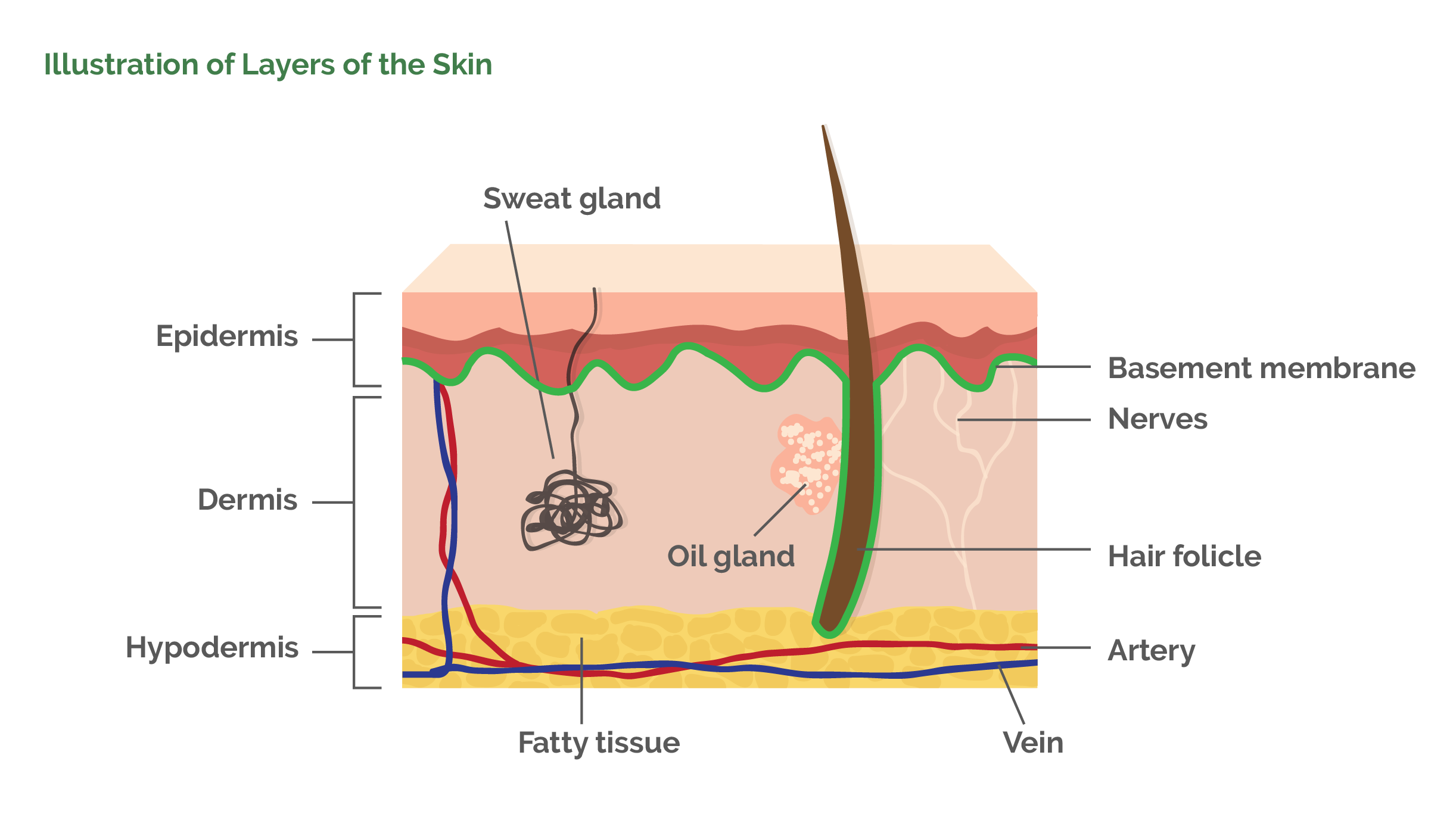
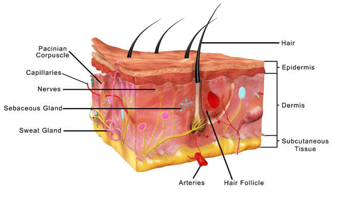
The Skin (Human Anatomy): Picture, Definition, Function, and ... Skin has three layers: The epidermis, the outermost layer of skin, provides a waterproof barrier and creates our skin tone. The dermis, beneath the epidermis, contains tough connective... Integumentary system parts: Quizzes and diagrams | Kenhub Sep 14, 2022 · Click below to download a free unlabeled version of the diagram above. Download PDF Worksheet (blank) Download PDF Worksheet (labeled) Skin anatomy. What if you want to test your knowledge of the skin only? No problem! With multiple layers and sublayers, there’s plenty to learn about skin anatomy. Check out our skin anatomy quiz below.
Skin Diagram with Detailed Illustrations and Clear Labels Skin Diagram with Detailed Illustrations and Clear Labels Biology Important Diagrams Skin Diagram Skin Diagram The largest organ in the human body is the skin, covering a total area of about 1.8 square meters. The skin is tasked with protecting our body from external elements as well as microbes. Interesting Note:
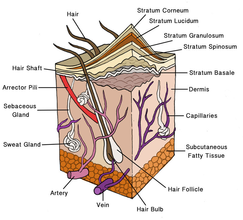
Skin diagram with labels
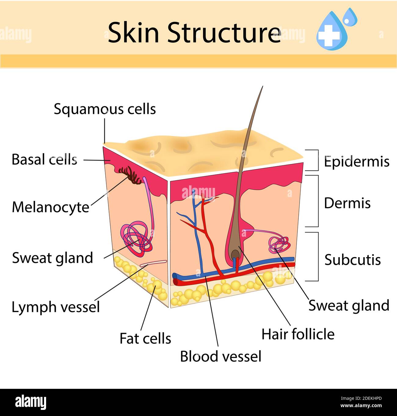
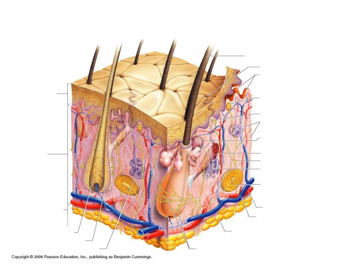
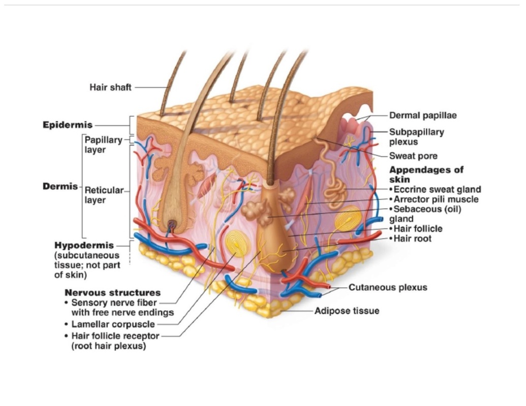
Skin Layers: Structure, Function, Anatomy, and More Mar 20, 2023 · There are three main layers of skin: Epidermis: The outermost layer, which contains five sub-layers. Dermis: The middle layer, which consists of two parts known as the papillary dermis (thin, upper layer) and the reticular dermis (thick, lower layer) Subcutaneous tissue: The deepest layer of skin. Labeled Skin Structure Diagram | Quizlet Skin Structure labeling 19 terms Diagram clarkson1994 Labeled Skin Structure 26 terms Images Diagram Ellen_Stanton8 Teacher Crash Course Nervous System Part 3 - Synapses 23 terms Julie_R_Schultz Teacher Bones of hand - anterior view 13 terms Diagram jpmartin971 Recent flashcard sets History 9weeks test 20 terms cepoling Special Test (4) 5.1 Layers of the Skin – Anatomy & Physiology Figure 5.1.1 – Layers of Skin: The skin is composed of two main layers: the epidermis, made of closely packed epithelial cells, and the dermis, made of dense, irregular connective tissue that houses blood vessels, hair follicles, sweat glands, and other structures.
Skin diagram with labels. Skin Model (labeled) Diagram | Quizlet Anatomy Skin Model (labeled) 5.0 (2 reviews) + − Flashcards Learn Test Match Created by Jacob_Curtis99 Terms in this set (11) Term epidermis Definition Top Layer Location Term Dermis Definition Middle Layer Location Term Hypodermis Definition Bottom Layer. (adipose tissue) Location Term Sebaceous Gland Location Term Arrector Pilli Muscle Location 5.1 Layers of the Skin – Anatomy & Physiology Figure 5.1.1 – Layers of Skin: The skin is composed of two main layers: the epidermis, made of closely packed epithelial cells, and the dermis, made of dense, irregular connective tissue that houses blood vessels, hair follicles, sweat glands, and other structures. Labeled Skin Structure Diagram | Quizlet Skin Structure labeling 19 terms Diagram clarkson1994 Labeled Skin Structure 26 terms Images Diagram Ellen_Stanton8 Teacher Crash Course Nervous System Part 3 - Synapses 23 terms Julie_R_Schultz Teacher Bones of hand - anterior view 13 terms Diagram jpmartin971 Recent flashcard sets History 9weeks test 20 terms cepoling Special Test (4) Skin Layers: Structure, Function, Anatomy, and More Mar 20, 2023 · There are three main layers of skin: Epidermis: The outermost layer, which contains five sub-layers. Dermis: The middle layer, which consists of two parts known as the papillary dermis (thin, upper layer) and the reticular dermis (thick, lower layer) Subcutaneous tissue: The deepest layer of skin.
Human Biology fig. 1.24 - Layers of the skin - English labels ...
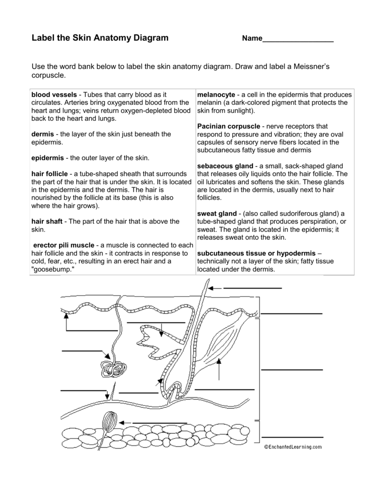
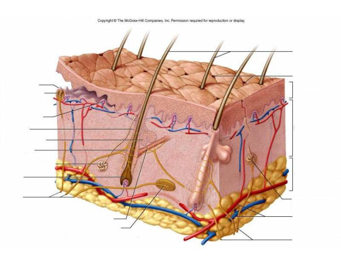
Label the Skin Anatomy Diagram

Typical skin structure diagram (Hill 2020). | Download ...
Human skin diagram hi-res stock photography and images - Alamy

Labeled Skin Structure Diagram | Quizlet

A diagrammatic representation of the structure of human skin ...
(118).jpg)
A Human Body Skin-structure Quiz! - ProProfs Quiz
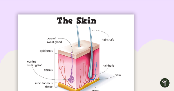
Skin Diagram Poster | Teach Starter
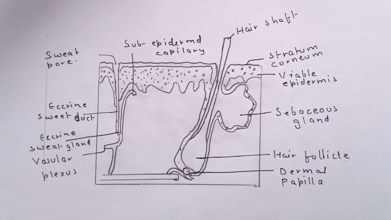
how to draw the diagram of human skin easily

The skin - 2023

Structure and Function of Skin | Biology for Majors II
skin anatomy Quiz

File:Skin.png - Wikimedia Commons
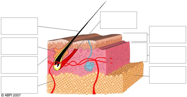
Quiz - Skin - ABPI - Resources for Schools

skin labeling Diagram | Quizlet
Anatomy of the Skin - Dermatology Sydney

Video 'Anatomy of a skin' A3 | Sylvia's H810 Online resource

Cross Section Human Skin Without Labels Stock Illustration ...

Schematic representation of basic human skin anatomy ...
Skin Cancers/ Dr Roger Graham: Plastic Surgeon/ Cape Town

Gen A&P, lab 10 the skin, skin diagram Diagram | Quizlet
Unit 4: Integumentary System - Badger Anatomy & Physiology

Human skin - Wikipedia

Anatomy Of Human Skin With Labels High-Res Vector Graphic ...

Skin Diagram Hair Shaft Stratum corneum Epidermis Stratum basale

Draw a Labelled Diagram to Show the Structure of Skin ...
Skin Layers Diagram Images – Browse 2,076 Stock Photos ...
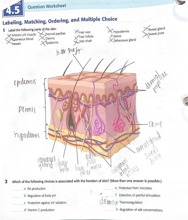
Solved 4.5 Label the following parts of the skin ***Which of ...
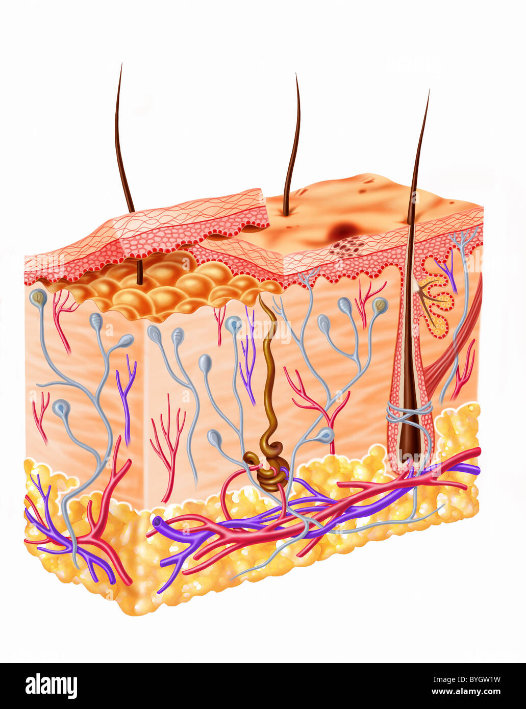
Human skin diagram hi-res stock photography and images - Alamy

File:Labeled layers of the skin.jpg - Wikipedia

Human Skin Anatomy, Labeled Version Stock Vector ...
Skin Labeling Quiz
The Skin - iQ Medical
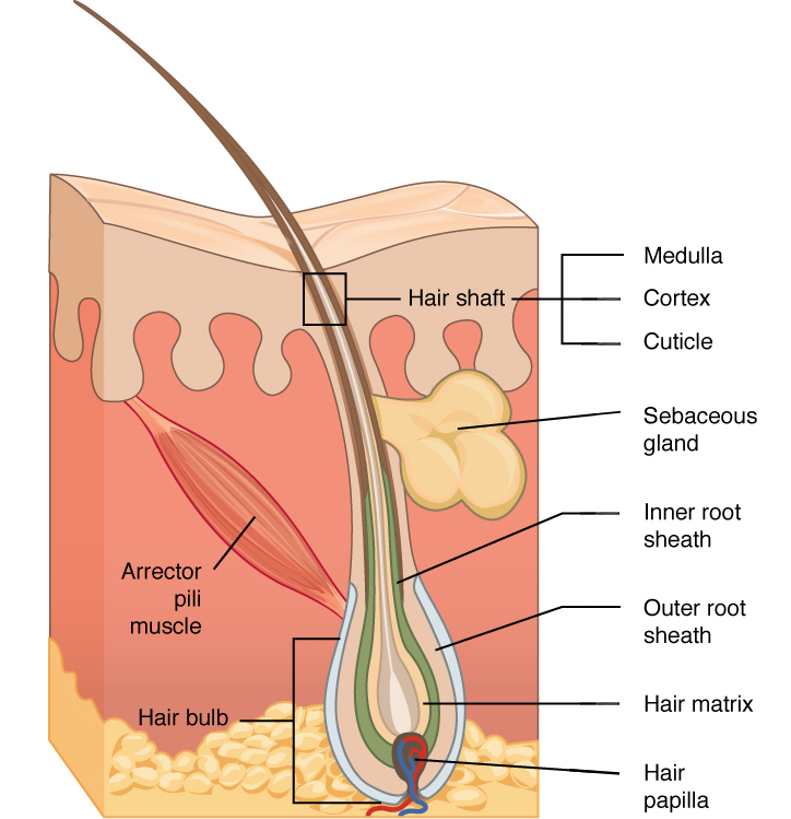
Hair | Biology for Majors II

Human Skin Diagram copy_resize | Pltch London | Flickr
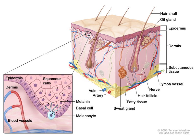
Skin: Image Details - NCI Visuals Online

protect yours.... | Anatomía de la piel, Psoriasis, El acne

Skin Diagram Worksheet - Have Fun Teaching
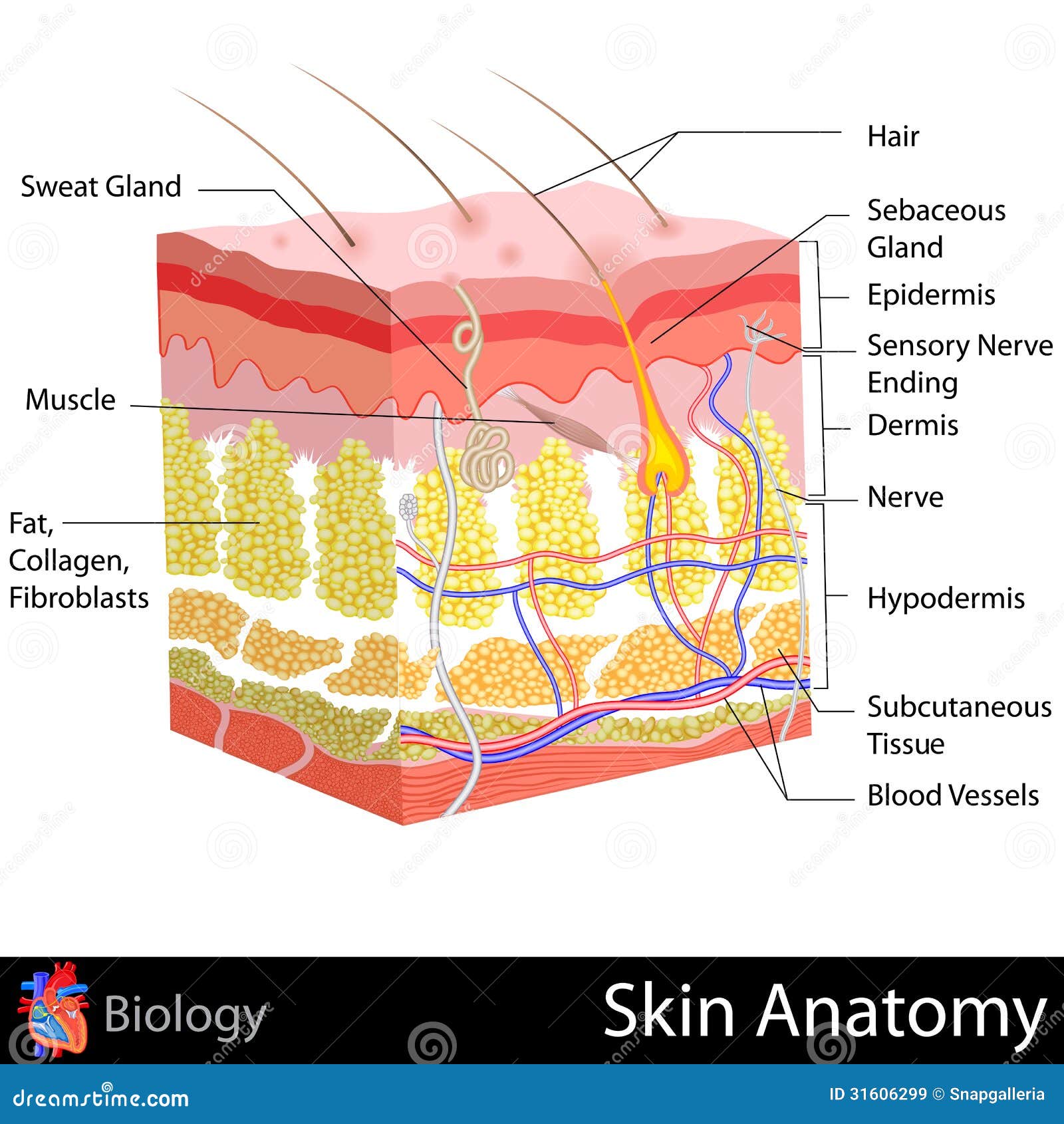
Skin Anatomy stock illustration. Illustration of layer - 31606299

Given below is a diagrammatic sketch of the vertical section ...
Post a Comment for "40 skin diagram with labels"